
Abstract
The background induced by the high penetration power of the  radiation is the main limiting factor of the current radio-guided surgery (RGS). To partially mitigate it, a RGS with
  probes less effective. We developed a
Introduction
The radio-guided surgery (RGS) is a surgical technique, first developed some 60 years ago, that enables the surgeon to evaluate the completeness of the tumoural lesion resection, while minimizing the amount of healthy tissue removed1. The impact of the RGS on the surgical management of cancer patients includes providing the surgeon with vital and real-time information regarding the location and the extent of the lesion, as well as assessing surgical resection margins. The technique (see Fig. 1) makes use of a radio-labelled tracer, preferentially taken up by the tumour to mark the cancerous tissue from the healthy organs and a probe (for a review see2), sensitive to the emission released by the tracer, to identify in real time the targeted tumour loci. The radio-pharmaceutical is administered to the patient just before the surgery.

The radioguided surgery technique (RGS).
Steps of the procedure: (1) a radio-labelled tracer is administered to the patient, before the surgery; (2) the emitting tracer is preferentially taken up by the tumour; (3) after the cancerous bulk removal, the surgeon explores the lesion with a radiation detecting probe and looks for targeted tumour residuals in real time. The bottom boxes show the effect of the proposed replacement of the  -emitting tracers (a) with electron-emitting tracers (b). Due to the high penetration power of the photons, in the first case a non-negligible background can be produced by the healthy organs close to the lesion, sometimes preventing the applicability of the technique. To mitigate this effect a shielding or active veto is applied (see inset of box a) thus making the probes cumbersome. Electrons, instead, provide a clearer delineation of radioactive tissue's margins allowing for a simple and compact probe and requiring a smaller radio-pharmaceutical activity. [Figure drawn by S.M. and E.S.C.].
Current clinical applications of the RGS are: radio-immuno-guided surgery (RIGS) for colon cancer, complete sentinel-node mapping for malignant melanoma and breast cancer and detection of parathyroid adenoma and bone tumours (such as osteoid osteoma).
Established methods make use of a combination of a  -emitting tracer with a
  radiation detection probe (see3 and references therein). Since
  radiation can traverse large amounts of tissue, any uptake of the tracer in nearby healthy tissue represents a non-negligible background, often preventing the usage of this technique.
To mitigate this effect it was suggested in literature the use of  with an energy of 511 keV: the background persists and actually increases in energy. The improvement with respect to the use of pure
  emitters is that a dual system can be devised where the background can be measured separately and subtracted from the observed signal. This approach has been studied in preclinical tests8,9,10,11,12 but it is not yet in use in the clinical practice. The largest limitations range from the time needed to identify a residual, the dimensions of the probes and the dose absorbed by the medical personnel.
A better solution to the current limits of RGS would be to eliminate the background from  radiation. This study suggests therefore the use of pure
  contamination, being the bremsstrahlung contribution, with a 0.1% emission probability, negligible. Furthermore, a
The idea of detecting
In particular, we report on the developments in the field of the radio-tracers and in the development and test of a specific intra-operative probe device that make the RGS with
Results
The clinical case
There are several possible applications of this technique, many of which probably have not yet been thought of. First, complete removal of tumour is critical for brain tumours, where a relapse is particularly dangerous and where other RGS techniques are limited by the high uptake of the brain. Next, particular attention should be paid to the complete resection of the main tumour and of infected lymph-nodes in the case of pediatric tumours where life expectancy is long. Such tumours are typically abdominal and therefore probe signals are blinded by background from kidneys, liver and bladder. Finally, there are abdominal tumours in adults, like non-palpable metastases in liver and insulinoma, that would profit from RGS.
Testing this novel technique with most of the above-mentioned clinical cases requires first to identify and test an appropriate
The β − detecting probe
As far as the probe design is concerned, the choice of the materials and the readout electronics is driven by the need to maximize the sensitivity to electrons while minimizing the sensitivity to photons. Para-terphenyl, or p-terphenyl, was adopted as electron detector after a detailed study18 due to its high light yield and low density, with consequent low sensitivity to photons.
A first prototype of the

First prototype of the intraoperative
The core is a cylindrical scintillator (diameter 2.1 mm, height 1.7 mm) of poli-crystalline p-terphenyl. A ring of PVC wraps the scintillator and shields it against radiation coming from the sides. The device is encapsulated inside an easy-to-handle aluminum body as protection against mechanical stress and it is protected against light by a thin PVC layer.
We explored the first prototype response with four phantoms simulating cancerous residuals with different topologies (see Supplementary Fig. S1). The 0.1 ml volume phantom, referred to as “RESIDUAL”, has dimensions compatible with residuals well identified with the nuclear magnetic resonance. To check the effect of the phantom depth on the probe response and resolution in distinguishing the residual edge, the other three cylindrical phantoms have the same activity concentration, footprint (13 mm2) but different heights: 1, 2 and 3 mm referred to as “H1”, “H2”, “H3” respectively.
A 1.5 mm step blind automated scan over the phantom engraved surface was performed when the activity concentration reached 16 kBq/ml. Even by limiting the acquisition time to 1 s per step the test demonstrated that all the residuals would be identifiable in absence of background from nearby organs. The signal from the tumor residuals was studied at maximum and minimum activity concentrations, 22 and 5 kBq/ml and the observed rates are reported in Tab. 1. The electronic noise was found to be negligible. Background rates, dominated by the signal coming from the uptake of nearby healthy tissue, were estimated using a full simulation of the system with FLUKA program19, based on the above mentioned DICOM images showing that the nearby healthy tissue uptake is approximately 10 times smaller than the tumour.
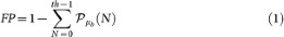
From the signal and background rates, taking into account the Poisson fluctuations of the measured counts, we computed the false-positive (FP) and the false-negative (FN) rates for a given time interval measurement with the probe. We estimated (see Tab. 1) the time needed to achieve FP≈ 1% and FN < 5% and found it to be 1 s for 22 kBq/ml activity concentration and up to 10 s in the case of a concentration as low as 5 kBq/ml. Further improvements of the probe are needed to reduce such time at a level compatible with surgical environment and administer down to 5 kBq/ml.
The effect of an off-axis measurement has been verified by moving the probe away from the phantom centre. The test showed that the rate is reduced by a factor 2 when moving the probe 0.5 mm far from the phantom edge (see Supplementary Fig. S4). This allows us to conclude that the probe distinguishes the residual edge. This test also demonstrated the lateral shielding effectiveness of this prototype making the probe insensitive to electrons coming from the sides hence increasing the tumor-spotting capability. This test also shows that the probe is insensitive of long-range radiation that could be caused by the activity of the 90Y in nearby samples.
Radiation exposure to patient and medical personnel
One of the key points of this technique is the low irradiation of the patient and above all of the medical staff. As previously mentioned, we estimated that a 22 kBq/ml activity concentration, corresponding to administering approximately 3 MBq/kg, would be more than sufficient for the technique to be effective.
From Ref. 16 we obtained that the total body dose absorbed by a 70 kg patient after administration of 210 MBq of 90Y-DOTATOC is approximately 21 mSv, while the effective dose is about 70 mSv. This corresponds to approximately two Computed Tomography (CT) exams and therefore there is room for improvement in reducing the required activity.
To evaluate the dose absorbed by the surgeons we simulated a set-up similar to the common situation of an operating room. Both activities of neoplastic cells and normal tissue were taken into account, according to the ratios obtained from the aforementioned studies on PET images. The equivalent dose absorbed by the surgeon's hands computed for RGS with a 90Y emitting tracer is expected to be smaller than 1
Discussion
The radio-guided surgery represents a very useful surgical adjunct in those cases where a complete resection, intended as full enhancing mass removal, is crucial both for recurrence-free survival and the overall survival of the patients, particularly for those tumours where the surgical mass removal is the only possible therapy.
We are proposing a radical change in the paradigm of this technique: the use of  or
or
This novel approach allows to develop a simple and compact probe which, detecting electrons and operating with low radiation background, provides a clearer delineation of margins of radioactive tissue and requires a smaller radio-pharmaceutical activity to detect tumour remnants compared to traditional RGS approaches. Moreover, due to the lower absorbed dose and the short range of electrons, the radiation exposure to the patient and medical personnel becomes almost negligible. Such considerations apply also in the comparison with the  background is in any case present, albeit subtracted with the dual-probe approach. Furthermore, measurements performed in absence of background, as in the case of the proposed
We have presented pre-clinical tests of a prototype probe supporting the above statements. From these measurements we extrapolated with a detailed simulation the expected performance with meningioma, representing our test clinical case. Nonetheless, the actual uptake on the margins of the lesion, the impact of tissue between the probe and the residual and the effect of the nearby blood is something to be estimated in clinical tests. Once the feasibility of such technique will be demonstrated with meningioma, it will be possible to extend it to other clinically relevant cases, eventually together with the development of specific radio-tracers.
Methods
The probe prototype
The core of the probe (see Fig. 2) is a cylindrical scintillator (diameter 2.1 mm, height 1.7 mm) of poli-crystalline para-terphenyl doped by 0.1% in mass of diphenylbutadiene. Para-terphenyl was adopted, after a detailed study18, due to its high light yield (3.5 times larger than typical organic scintillators), non-hygroscopic property and low density. The scintillator is shielding against radiation coming from the sides by wrapping it with a 7 mm external diameter ring of PVC. The device is encapsulated inside an easy-to-handle aluminum body, as protection against mechanical stress and has a blinding 0.4 mm-thick black PVC front end cap. The scintillation light is transported to a photo-multiplier tube (PMT, Hamamatsu H10721-210), through an optical fibre and read out by a portable custom electronics with wireless connection to a PC or tablet. This prototype is compatible with a standard sterile covering of sub-millimetric film for surgical environment. Also the very low bias required by the PMT, 5 V, makes the device easily portable.
Experimental set-up of the 90Y test
The phantoms are cylindrical vessels, engraved at a fixed radius on a PMMA (Plexiglass™) disk mounted on a rotating table, as shown in supplementary Fig. S1. Their dimensions are listed in Tab. 1 and are known with a precision of 10
During the test, the probe was fixed in vertical position over the disk and the phantoms were moved under the probe by rotating the disk with a step motor, controlled via PC. The relative position of the sensitive head of the probe and the phantom was known with a precision of 10
Estimate of false positive and false negative rates
The rate of false positive and false negative (FP and FN) is computed from the signal (
where  indicates the Poisson probability to have N if the mean is
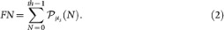
To determine the minimum acquisition time reported in Tab. 1, FN and FP are computed in a grid of tdaq and th (see Supplementary Fig. S5) and the smallest value of tdaq for which FN < 5% and FP≈ 1% is determined.
References
Radioguided Surgery: A Comprehensive Team Approach. [Mariani, G., Giuliano, A. E. & Strauss, H. W. (eds)] (Springer, New York, 2006).
Hoffman, E. J. Tornai, M. P. Janecek, M. Patt, B. E. & Iwanczyk, J. S. Intraoperative probes and imaging probes. Eur. J. Nucl. Med. 26, 913–935 (1999).
Tsuchimochi, M. & Hayamaand, K. Intraoperative gamma cameras for radioguided surgery: Technical characteristics, performance parameters and clinical application. Phys. Med. 29, 126–38 (2013).
Hickernell, T. S. et al. Dual detector Probe for surgical Tumor Staging. J. Nucl. Med. 29, 1101 (1988).
Daghighian, F. et al. Intraoperative beta probe: A device for detecting tissue labeled with positron or electron emitting isotopes during surgery. Med. Phys. 21, 153 (1994).
Raylman, R. R. & Wahl, R. L. A fiber-optically coupled positron-sensitive surgical probe. J. Nucl. Med. 35, 909 (1994).
Bonzom, S. An Intraoperative Beta Probe Dedicated to Glioma Surgery: Design and Feasibility Study. IEEE Trans. Nucl. Sci. 54, 1 (2007).
Raylman, R. R. et al. Fluorine-18-fluorodeoxyglucose-guided breast cancer surgery with a positron-sensitive probe: Validation in preclinical studies. J. Nucl. Med. 36, 1869 (1995).
Zervos, E. E. et al. 18F-Labeled Fluorodeoxyglucose Positron Emission Tomography-Guided Surgery for Recurrent Colorectal Cancer: A Feasibility Study. J. of Surg. Res. 97, 9 (2001).
Bogalhas, F. et al. Development of a positron probe for localization and excision of brain tumours during surgery. Phys. Med. Biol. 54, 4439 (2009).
Gonzales, S. J. et al. An analysis of the utility of handheld PET probes for the intraoperative localization of malignant tissue. J. Gastrointest. Surg. 15, 358–55 (2011).
Singh, B. et al. A hand-held beta imaging probe for FDG. Ann. of Nucl. Med. 27, 203–208 (2013).
Selverstone, B., Solomon, A. K. & Sweet, W. H. Location of brain tumors by means of radioactive phosphorous. The J. of the American Med. Ass. 140, 277–278 (1949).
Cremonesi, M. Ferrari, M. Bodei, L. Tosi, G. & Paganelli, G. Dosimetry in peptide radionuclide receptor therapy: a review. J. Nucl. Med. 47, 1467–1475 (2006).
Bartolomei, M. et al. Peptide receptor radionuclide therapy with 90Y-DOTATOC in recurrent meningioma. Eur. J. Nucl. Med. Mol. Imaging 36, 1407–1416 (2009).
Cremonesi, M. et al. Dosimetry for treatment with radiolabelled somatostatin analogues. A review. Q. J. Nucl. Med. Mol. Imaging 54, 37–51 (2010).
Fabbri, C. et al. Quantitative evaluation on 90Y–DOTATOC PET and SPECT imaging by phantom acquisitions and clinical applications in locoregional and systemic treatments. Q. J. Nucl. Med. Mol. Imaging 56, 522–8 (2012).
Angelone, M. et al. Properties of para-terphenyl as detector for alpha, beta and gamma radiation. arXiv:1305.0442 (2013).
Battistoni, G. et al. The FLUKA code: Description and benchmarking. AIP Conf. Proc. 896, 31–49 (2006).
Acknowledgements
We would like to thank the Institute for Nuclear Medicine of the Policlinico A. Gemelli, Universita' Cattolica del Sacro Cuore of Rome for the stimulating discussions.
Author information
Authors and Affiliations
Contributions
R.F., A.Sc. and S.F. ideated the project and supervise it by identifying the materials and their supply; S.M., E.S.C., A.R., F.C., F.B., M.M. and A.Sa. worked on the design and construction of the probe prototype, automated test station implementation, data analysis and interpretation, probe optimization; R.F., E.S.C., S.M. and F.C. prepared the manuscript; F.C., V.P., L.P., I.M., C.V. and E.D.L. implemented the Monte Carlo simulation aimed to compare different materials and to understand the probe behavior; G.B., P.F. and M.S. provided the medical background and defined the clinical case; G.P., C.M.G. and M.C. provided the nuclear medicine knowledge and supplied DICOM data to study use cases; V.B. and L.R. developed and optimized custom electronics for the probe readout; all authors discussed the results and implications and commented on the manuscript at all stages.
Ethics declarations
Competing interests
F.B., F.C., E.D.L., S.F., M.M., I.M., V.P., L.P., A.Sa., A.Sc., C.V. and R.F. are listed as inventors on an Italian patent application (RM2013A000053) entitled “Utilizzo di radiazione
Electronic supplementary material
Supplementary Information
A novel radioguided surgery technique exploiting beta- decays
Rights and permissions
This work is licensed under a Creative Commons Attribution-NonCommercial-NoDerivs 3.0 Unported License. To view a copy of this license, visit http://creativecommons.org/licenses/by-nc-nd/3.0/
About this article
Cite this article
Camillocci, E., Baroni, G., Bellini, F. et al. A novel radioguided surgery technique exploiting
Received:
Accepted:
Published:
DOI: https://doi.org/10.1038/srep04401
