Category:Amnion
Jump to navigation
Jump to search
innermost membranous sac that surrounds and protects the developing embryo  | |||||
| Upload media | |||||
| Instance of |
| ||||
|---|---|---|---|---|---|
| Subclass of |
| ||||
| |||||
Media in category "Amnion"
The following 70 files are in this category, out of 70 total.
-
 2913 Embryonic Folding.jpg 1,894 × 1,735; 831 KB
-
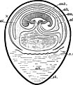
 A human embryo of 2 mm. in median sagittal section.jpg 838 × 1,006; 310 KB
-

-

-
 Allantois bird.jpg 1,062 × 1,674; 366 KB
-
 Amnion formation in mouse embryos, illustrated by longitudinal sections.jpg 1,200 × 1,394; 1.37 MB
-
 Amnion Formation In Mouse Embryos, Illustrated By Transverse Sections.jpg 1,200 × 1,416; 969 KB
-
 Amniote embryo ku.jpg 1,162 × 871; 206 KB
-
 Amniote embryo.jpg 1,268 × 954; 415 KB
-

-
 Aves Embryo of aboul 27 somites drawn in alcohol by reflected light; upper side, x 10.jpg 1,287 × 1,385; 1.68 MB
-
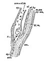
 Aves Median longitudinal section of a thirty-six-hour chick embryo.jpg 833 × 1,729; 271 KB
-

-
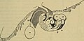
 Aves Transverse sections through embryo fifth day after incubation.jpg 688 × 755; 357 KB
-
 Baby gives birth to Tia.jpg 2,048 × 1,536; 1.38 MB
-
 Calopteryx embryo development.jpg 669 × 740; 429 KB
-
 Catfetus1.jpg 1,082 × 1,372; 165 KB
-
 Chicken embryo of about five days incubation.jpg 1,218 × 750; 1,003 KB
-
 Chicken embryo of about fourteen days incubation.jpg 1,216 × 736; 901 KB
-
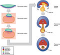
 Development of the amnion and allantois.jpg 676 × 902; 1.05 MB
-
 Diagrams and images of human embryos at the gastrula stage.png 3,128 × 3,193; 804 KB
-
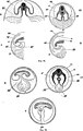
 Diagrams showing the development of the amnion, chorion and allantois.jpg 1,269 × 1,151; 605 KB
-
 Embryonic and extraembryonic ectoderm demarcation in the amniochorionic fold.jpg 1,200 × 1,937; 1.62 MB
-
 Extra-embryonic membranes of the chic (01).jpg 641 × 783; 558 KB
-
 Extra-embryonic membranes of the chick.jpg 897 × 455; 438 KB
-
 Extraembryonic tissues and organs in a mouse embryo and foetus.jpg 1,200 × 581; 542 KB
-
 Foetus cat (01).jpg 954 × 612; 497 KB
-
 Foetus cat.jpg 667 × 827; 532 KB
-
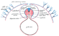
 Formation of the Umbilicus and Allantois. human embryo, 0.7 mm. long..jpg 1,152 × 803; 970 KB
-
 Formation- of the Umbilicus in an Embryo 2.5 mm.jpg 788 × 837; 598 KB
-
 Four diagrams showing hypothetical stages of early human embryos.jpg 1,631 × 1,434; 943 KB
-
 Fowl embryo (01).jpg 768 × 1,021; 246 KB
-
 Fowl embryo.jpg 556 × 640; 131 KB
-
 Gray12.png 1,115 × 491; 192 KB
-
 Gray22.png 300 × 303; 23 KB
-
 Gray29.png 500 × 306; 15 KB
-
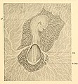
 Hand-book of physiology (1892) (14785304043).jpg 1,792 × 888; 162 KB
-
 Human embryo Section of embryonic rudiment in Peters' ovum (first week).jpg 1,141 × 857; 540 KB
-
 Human- Embryo, about 3.5 mm. long.jpg 803 × 791; 701 KB
-
 Human- Embryo, about 5 mm. long.jpg 825 × 846; 809 KB
-
 Hydrophilus piceus embryos (01).jpg 754 × 750; 520 KB
-
 Hydrophilus piceus embryos.jpg 815 × 657; 616 KB
-
 Insect development of the embryonic envelopes.jpg 980 × 629; 614 KB
-

-

-
 Morphological differences between human and mouse gastrulation.jpg 2,994 × 3,411; 397 KB
-

-
 Pig embryo median sagittal section.jpg 1,257 × 1,143; 617 KB
-
 Pig embryo transverse section.jpg 889 × 772; 397 KB
-
 Reconstruction of embryos prepared for kaufman's the atlas of mouse development.jpg 1,220 × 2,124; 1.09 MB
-
 Sagittal Section of Human Zygote.jpg 817 × 684; 653 KB
-

-

-

-

-
 Section showing three stages in the formation of the amnion of bat embryo.jpg 1,407 × 1,695; 911 KB
-
 Series of longitudinal sections of an embryo with large exocoelomic cavity (ec).jpg 1,220 × 1,217; 1.12 MB
-
 Simiiformes developing blastocyst.jpg 1,105 × 1,369; 668 KB
-

-
 Structure of the human amniotic membrane.jpg 3,313 × 3,966; 832 KB
-
 Text-book of embryology (1914) (20153062293).jpg 1,306 × 1,972; 475 KB
-
 The development of the chick - an introduction to embryology (1936) (20703235188).jpg 1,219 × 1,645; 1.56 MB
-
 The development of the chick; an introduction to embryology (1908) (20881489692).jpg 1,868 × 1,940; 1.26 MB
-
 The development of the chick; an introduction to embryology (1919) (14755464245).jpg 1,934 × 2,704; 1,008 KB
-
 Three-dimensional analyses of cloacal division processes.jpg 1,575 × 1,368; 1.35 MB
-

-

-
 Umbilical Cord of a Human Embryo 12.5 mm. in length.jpg 1,001 × 1,445; 1.15 MB
-
 Umbilical Region of a Human Embryo 10 mm. in length.jpg 1,167 × 957; 1.09 MB
-
 Yolk sacs.png 1,346 × 511; 294 KB


.jpg)


.jpg)


.jpg)


_(14785304043).jpg)
.jpg)


.jpg)


._Reichert%27s_membrane_omitted.jpg)


.jpg)


_(20153062293).jpg)
_(20703235188).jpg)
_(20881489692).jpg)
_(14755464245).jpg)


_in_correspondence_of_the_chorion_laeve_(hCL)_and_the_capsular_decidua_(hCD).jpg)