Category:Electron diffraction
Jump to navigation
Jump to search
Bending of electron beams due to electrostatic interactions with matter | |||||
| Upload media | |||||
| Subclass of | |||||
|---|---|---|---|---|---|
| |||||
Subcategories
This category has the following 8 subcategories, out of 8 total.
Media in category "Electron diffraction"
The following 52 files are in this category, out of 52 total.
-
 A565 G P Thomson Electron Diffraction.jpg 1,525 × 1,536; 159 KB
-
 Al-Cu-Fe-Cr decagonal quasicrystal diffraction pattern.tif 1,273 × 1,281; 1.45 MB
-
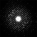
 Aluminumpowderpattern.png 512 × 488; 178 KB
-
 Bell Labs APS plaque west side of Westbeth door jeh edited.jpg 3,460 × 1,853; 2.4 MB
-
 CBED scheme.png 927 × 1,202; 23 KB
-
 CBED sketch.png 1,641 × 1,222; 118 KB
-
 CBED-EFiltered.png 1,182 × 919; 1.16 MB
-
 CBEDThickness.png 902 × 915; 668 KB
-
 Claus Jönsson Interferenz.jpg 146 × 200; 1 KB
-
 Crystal orientation and diffraction.gif 800 × 400; 2.13 MB
-
 Davisson-Germer experiment.svg 900 × 650; 3 KB
-
 Diff.jpg 168 × 172; 16 KB
-
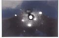
 Diffraction of electrons on microcrystallitic crystals or po Wellcome V0018969EBR.jpg 1,356 × 1,388; 884 KB
-
 Diffraction of electrons on microcrystallitic crystals or po Wellcome V0018969ETL.jpg 1,356 × 1,345; 900 KB
-
 Diffraction of electrons on microcrystallitic crystals or po Wellcome V0018969ETR.jpg 1,339 × 1,359; 933 KB
-
 DifraccionElectronesMET.jpg 354 × 354; 63 KB
-
 Difrakce.png 1,653 × 725; 294 KB
-
 Electron diffraction Laue-zone tilt-series.gif 360 × 360; 489 KB
-
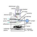
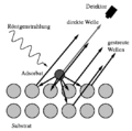
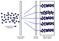
 Electron diffraction setup.svg 500 × 200; 86 KB
-
Electron Diffraction Tube Graphite.webm 6.0 s, 1,920 × 1,080; 197 KB
-
 Electron diffraction.svg 400 × 265; 26 KB
-
 Electron gun jyu.jpg 700 × 467; 105 KB
-
 Electron scattering.png 960 × 720; 10 KB
-
 ElectronDiffraction01.jpg 3,020 × 2,456; 1.59 MB
-
 ElectronDiffraction02.jpg 2,584 × 1,720; 1,007 KB
-
 ElectronDiffraction03.jpg 2,636 × 2,176; 1.36 MB
-
 ElectronDiffraction04.jpg 3,148 × 2,436; 1.67 MB
-
 Elektronenbeugung - DS7 6133 PK.jpg 2,172 × 2,410; 2.39 MB
-
 Em diffraction.jpg 167 × 125; 23 KB
-
 Ewald3.png 1,146 × 1,047; 127 KB
-
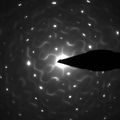
 GED C6H6 diff pattern.jpg 756 × 756; 37 KB
-
 GED scheme 1.jpg 756 × 756; 87 KB
-
 Incomm.png 584 × 546; 319 KB
-
 KMapFCC.png 1,252 × 1,010; 602 KB
-
 LEED pattern of CO on Platinum-Rhodium (100) surface, E = 94 eV.JPG 3,609 × 2,377; 1.4 MB
-

-
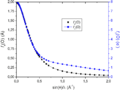
 NbCoSb showing diffuse scattering.png 1,072 × 1,072; 451 KB
-
 Oxygen form factor - e & X.png 1,468 × 1,099; 22 KB
-
 Photoelektronenbeugung.png 529 × 523; 7 KB
-
 Rockingcurve2.png 370 × 336; 36 KB
-
 Roger Bach et al 2013 New J. Phys. 15 033018 Figure 3.jpg 324 × 813; 75 KB
-
 SADvsCBED.svg 982 × 845; 5 KB
-
 Si100Reconstructed.png 2,072 × 1,944; 5.31 MB
-
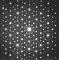
 Si111 7x7 ReconstructionB.png 512 × 286; 99 KB
-
 Simple scattering.gif 600 × 600; 241 KB
-
 Tant-ED.jpg 512 × 512; 117 KB
-
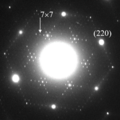 Transmission electron diffraction pattern of Si (111) 7x7.png 481 × 483; 411 KB
-
 Two-Slit Experiment Electrons.svg 500 × 350; 59 KB
-
 XtaLAB Synergy-ED by Rigaku.jpg 800 × 610; 18 KB
-
 Zn-Mg-HoDiffraction.JPG 364 × 372; 15 KB
-
 Дифракція повільних елекронів (ДПЕ) на монокристалічному молібдені.jpg 1,200 × 801; 436 KB
-
 Դեյվիսըն-Ջերմերի փորձ.svg 421 × 296; 22 KB


_surface,_E_%3D_94_eV.JPG)
_surface_-_STM_and_LEED_characterization.svg)


_%D0%BD%D0%B0_%D0%BC%D0%BE%D0%BD%D0%BE%D0%BA%D1%80%D0%B8%D1%81%D1%82%D0%B0%D0%BB%D1%96%D1%87%D0%BD%D0%BE%D0%BC%D1%83_%D0%BC%D0%BE%D0%BB%D1%96%D0%B1%D0%B4%D0%B5%D0%BD%D1%96.jpg)

_7x7.png)