Respiratory epithelium is a type of ciliated epithelium found lining most of the respiratory tract, where it serves to moisten and protect the airways. It is not present in the larynx and pharynx. It also functions as a barrier to potential pathogens and foreign particles, preventing infection and tissue injury by the action of mucociliary clearance.
| Respiratory epithelium | |
|---|---|
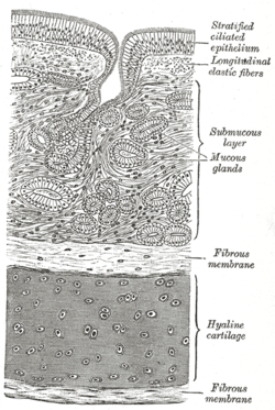
 Diagram of the trachea showing respiratory epithelium at the top | |
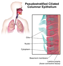
 Illustration depicting Pseudostratified Ciliated Columnar Epithelium. | |
| Details | |
| System | Respiratory system |
| Identifiers | |
| MeSH | D020545 |
| TH | H3.05.00.0.00003 |
| Anatomical terms of microanatomy | |
Structure
The respiratory epithelium lining the upper respiratory airways is classified as ciliated pseudostratified columnar epithelium.[1] This designation is due to the arrangement of the multiple cell types composing the respiratory epithelium. While all cells make contact with the basement membrane and are, therefore, a single layer of cells, the nuclei are not aligned in the same plane. Hence, it appears as though several layers of cells are present and the epithelium is called pseudostratified.
The majority of cells composing the ciliated pseudostratified columnar epithelium are of three types: a) ciliated cells, b) goblet cells, and c) basal cells. The ciliated cells are columnar epithelial cells with specialized ciliary modifications. Goblet cells, so named because they are shaped like a wine goblet, are columnar epithelial cells that contain membrane-bound mucous granules and secrete mucus, or epithelial lining fluid (ELF), the composition of which is tightly regulated; the mucus helps maintain epithelial moisture and traps particulate material and pathogens moving through the airway. and determines how well mucociliary clearance works.[2][3] The basal cells are small, nearly cuboidal cells thought to have some ability to differentiate into other cells types found within the epithelium. For example, these basal cells respond to injury of the airway epithelium, migrating to cover a site denuded of differentiated epithelial cells, and subsequently differentiating to restore a healthy epithelial cell layer.
Other cells of the respiratory epithelium include brush cells, which are columnar cells with microvilli that function as chemoreceptors, and small granule cells (Kulchitsky cells), which are neuroendocrine cells.[4]
Certain parts of the respiratory tract, such as the oropharynx, are also subject to the abrasive swallowing of food. To prevent the destruction of the respiratory epithelium in these areas, it changes to stratified squamous epithelium, which is better suited to the constant sloughing and abrasion. The squamous layer of the oropharynx is continuous with the esophagus.[citation needed]
References
- ^ Mescher AL, "Chapter 17. The Respiratory System" (Chapter). Mescher AL: Junqueira's Basic Histology: Text & Atlas, 12e: "Archived copy". Archived from the original on 2013-06-03. Retrieved 2015-02-24.
{{cite web}}: Unknown parameter|deadurl=ignored (|url-status=suggested) (help)CS1 maint: archived copy as title (link). - ^ Stanke F The Contribution of the Airway Epithelial Cell to Host Defense. Mediators Inflamm. 2015;2015:463016. PMID 26185361 PMC 4491388
- ^ U.S. EPA. Integrated Science Assessment for Oxides of Nitrogen – Health Criteria (2016 Final Report). U.S. Environmental Protection Agency, Washington, DC, EPA/600/R-15/068, 2016. Federal Register Notice Jan 28, 2016 Free download available at Report page at EPA website.
- ^ Histology: A Text and Atlas, with Correlated Cell and Molecular Biology, 7th Edition. p. 664.
External links
- Histology image: 13903loa – Histology Learning System at Boston University
- Histology of The Respiratory System