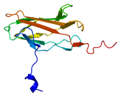
Runt-related transcription factor 3 is a protein that in humans is encoded by the RUNX3 gene.[5]
Function
editThis gene encodes a member of the runt domain-containing family of transcription factors. A heterodimer of this protein and a beta subunit forms a complex that binds to the core DNA sequence 5'-YGYGGT-3' found in a number of enhancers and promoters,[6] and can either activate or suppress transcription. It also interacts with other transcription factors. It functions as a tumor suppressor, and the gene is frequently deleted or transcriptionally silenced in cancer. Multiple transcript variants encoding different isoforms have been found for this gene.[7]
In melanocytic cells RUNX3 gene expression may be regulated by MITF.[8]
RUNX3 plays a fundamental role in defense against early tumor formation. In response to growth factors, RUNX3 is acetylated by p300 to complex with bromodomain-containing protein 2 (BRD2; a member of the BET family of transcription co-regulators)[9] and to subsequent transient induction of CDKN1A and ARF.[10] CDKN1A (also known as CIP1 or p21) inhibits the cell cycle, and ARF inhibits MDM2, increasing the stability of the cancer-suppressing gene p53.[10]
The expression of CDKN1A and ARF under wild-type cell cycles is temporary, which results from the RUNX3-BRD2 complex replacing the RUNX3-cyclinD1 complex. However, oncogenic mitogen signals such as KRASG12D cause the RUNX3-BRD2 complex to be maintained continuously, resulting in the continuous expression of p21, ARF, and p53. Therefore, RUNX3 can function as a sensor for unregulated mitogenic signals, and its inactivation can ultimately lead to cancer due to the loss of function as a sensor.[10]
Knockout mouse
editRunx3 null mouse gastric mucosa exhibits hyperplasia due to stimulated proliferation and suppressed apoptosis in epithelial cells, and the cells are resistant to TGF-beta stimulation.[11]
The RUNX3 controversy and resolution
editIn 2011 doubt was cast over the tumor suppressor function of Runx3 originated from the earlier publication by Li and co-workers.[12] On the basis of the original study by Li and co-workers (2002), the majority of later literature citing Li and co-workers (2002) assumed that RUNX3 was expressed in the normal gut epithelium and that it is therefore likely to act as a tumor suppressor in the particular epithelial cancer investigated. Most of this literature used RUNX3 promoter methylation status in various cancers as a proxy for its expression. However, quite many genes are known to be methylated in tumor cell genomes, and the majority of these genes are not expressed in the normal tissue of origin of these cancers. Others used poorly characterized (or fully invalidated) antibodies to detect the RUNX3 protein, or used RT-PCR or validated antibodies and failed to detect RUNX3 in the gut epithelium but still did not question the original finding by Li and co-workers (2002). This facts have recently been discussed in a book by Ülo Maiväli.[13]
In late 2009, a report written by Kosei Ito and his co-workers resolved the controversy by verifying that RUNX3 is indeed expressed in human and mouse gastrointestinal tract (GIT) epithelium and it functions as a tumor suppressor in gastric and colorectal tissues.[14] The authors of the paper suggested that the previous conflicting report might be caused by use of a specific antibody, known as G-poly. Ito and his team generated multiple anti-RUNX3 monoclonal antibodies recognizing the RUNX3 N-terminal region (residues 1-234). The researchers found that the antibodies react with RUNX3 in gastric epithelial cells, whereas those recognizing the C-terminal region did not. G-poly primarily recognizes the region beyond 234 and hence, is unable to detect Runx3 in this tissue.
Interactions
editSee also
editReferences
edit- ^ a b c GRCh38: Ensembl release 89: ENSG00000020633 – Ensembl, May 2017
- ^ a b c GRCm38: Ensembl release 89: ENSMUSG00000070691 – Ensembl, May 2017
- ^ "Human PubMed Reference:". National Center for Biotechnology Information, U.S. National Library of Medicine.
- ^ "Mouse PubMed Reference:". National Center for Biotechnology Information, U.S. National Library of Medicine.
- ^ Levanon D, Negreanu V, Bernstein Y, Bar-Am I, Avivi L, Groner Y (September 1994). "AML1, AML2, and AML3, the human members of the runt domain gene-family: cDNA structure, expression, and chromosomal localization". Genomics. 23 (2): 425–432. doi:10.1006/geno.1994.1519. PMID 7835892.
- ^ Levanon D, Eisenstein M, Groner Y (April 1998). "Site-directed mutagenesis supports a three-dimensional model of the runt domain". Journal of Molecular Biology. 277 (3): 509–512. doi:10.1006/jmbi.1998.1633. PMID 9533875. S2CID 13139512.
- ^ "Entrez Gene: RUNX3 runt-related transcription factor 3".
- ^ Hoek KS, Schlegel NC, Eichhoff OM, Widmer DS, Praetorius C, Einarsson SO, et al. (December 2008). "Novel MITF targets identified using a two-step DNA microarray strategy". Pigment Cell & Melanoma Research. 21 (6): 665–676. doi:10.1111/j.1755-148X.2008.00505.x. PMID 19067971.
- ^ Lee YS, Lee JW, Jang JW, Chi XZ, Kim JH, Li YH, et al. (November 2013). "Runx3 inactivation is a crucial early event in the development of lung adenocarcinoma". Cancer Cell. 24 (5): 603–616. doi:10.1016/j.ccr.2013.10.003. PMID 24229708.
- ^ a b c Lee JW, Kim DM, Jang JW, Park TG, Song SH, Lee YS, et al. (April 2019). "RUNX3 regulates cell cycle-dependent chromatin dynamics by functioning as a pioneer factor of the restriction-point". Nature Communications. 10 (1): 1897. Bibcode:2019NatCo..10.1897L. doi:10.1038/s41467-019-09810-w. PMC 6479060. PMID 31015486.
- ^ Li QL, Ito K, Sakakura C, Fukamachi H, Inoue K, Chi XZ, et al. (April 2002). "Causal relationship between the loss of RUNX3 expression and gastric cancer". Cell. 109 (1): 113–124. doi:10.1016/S0092-8674(02)00690-6. PMID 11955451. S2CID 11362226.
- ^ Levanon D, Bernstein Y, Negreanu V, Bone KR, Pozner A, Eilam R, et al. (October 2011). "Absence of Runx3 expression in normal gastrointestinal epithelium calls into question its tumour suppressor function". EMBO Molecular Medicine. 3 (10): 593–604. doi:10.1002/emmm.201100168. PMC 3258485. PMID 21786422.
- ^ Maiväli Ü (2015). Interpreting Biomedical Science. Academic Press. pp. 44–45. ISBN 9780124186897.
- ^ Ito K, Inoue KI, Bae SC, Ito Y (March 2009). "Runx3 expression in gastrointestinal tract epithelium: resolving the controversy". Oncogene. 28 (10): 1379–1384. doi:10.1038/onc.2008.496. PMID 19169278.
- ^ Levanon D, Goldstein RE, Bernstein Y, Tang H, Goldenberg D, Stifani S, et al. (September 1998). "Transcriptional repression by AML1 and LEF-1 is mediated by the TLE/Groucho corepressors". Proceedings of the National Academy of Sciences of the United States of America. 95 (20): 11590–11595. Bibcode:1998PNAS...9511590L. doi:10.1073/pnas.95.20.11590. PMC 21685. PMID 9751710.
Further reading
edit- Vogiatzi P, De Falco G, Claudio PP, Giordano A (April 2006). "How does the human RUNX3 gene induce apoptosis in gastric cancer? Latest data, reflections and reactions". Cancer Biology & Therapy. 5 (4): 371–374. doi:10.4161/cbt.5.4.2748. PMID 16627973.
- Wijmenga C, Speck NA, Dracopoli NC, Hofker MH, Liu P, Collins FS (April 1995). "Identification of a new murine runt domain-containing gene, Cbfa3, and localization of the human homolog, CBFA3, to chromosome 1p35-pter". Genomics. 26 (3): 611–614. doi:10.1016/0888-7543(95)80185-O. PMID 7607690.
- Bae SC, Takahashi E, Zhang YW, Ogawa E, Shigesada K, Namba Y, et al. (July 1995). "Cloning, mapping and expression of PEBP2 alpha C, a third gene encoding the mammalian Runt domain". Gene. 159 (2): 245–248. doi:10.1016/0378-1119(95)00060-J. PMID 7622058.
- Bae SC, Yamaguchi-Iwai Y, Ogawa E, Maruyama M, Inuzuka M, Kagoshima H, et al. (March 1993). "Isolation of PEBP2 alpha B cDNA representing the mouse homolog of human acute myeloid leukemia gene, AML1". Oncogene. 8 (3): 809–814. PMID 8437866.
- Levanon D, Goldstein RE, Bernstein Y, Tang H, Goldenberg D, Stifani S, et al. (September 1998). "Transcriptional repression by AML1 and LEF-1 is mediated by the TLE/Groucho corepressors". Proceedings of the National Academy of Sciences of the United States of America. 95 (20): 11590–11595. Bibcode:1998PNAS...9511590L. doi:10.1073/pnas.95.20.11590. PMC 21685. PMID 9751710.
- Bangsow C, Rubins N, Glusman G, Bernstein Y, Negreanu V, Goldenberg D, et al. (November 2001). "The RUNX3 gene--sequence, structure and regulated expression". Gene. 279 (2): 221–232. doi:10.1016/S0378-1119(01)00760-0. PMID 11733147.
- Waki T, Tamura G, Sato M, Terashima M, Nishizuka S, Motoyama T (April 2003). "Promoter methylation status of DAP-kinase and RUNX3 genes in neoplastic and non-neoplastic gastric epithelia". Cancer Science. 94 (4): 360–364. doi:10.1111/j.1349-7006.2003.tb01447.x. PMC 11160204. PMID 12824905. S2CID 22030810.
- Puig-Kröger A, Sanchez-Elsner T, Ruiz N, Andreu EJ, Prosper F, Jensen UB, et al. (November 2003). "RUNX/AML and C/EBP factors regulate CD11a integrin expression in myeloid cells through overlapping regulatory elements". Blood. 102 (9): 3252–3261. doi:10.1182/blood-2003-02-0618. hdl:10171/18733. PMID 12855590.
- Kato N, Tamura G, Fukase M, Shibuya H, Motoyama T (August 2003). "Hypermethylation of the RUNX3 gene promoter in testicular yolk sac tumor of infants". The American Journal of Pathology. 163 (2): 387–391. doi:10.1016/S0002-9440(10)63668-1. PMC 1868235. PMID 12875960.
- Yang N, Zhang L, Zhang Y, Kazazian HH (August 2003). "An important role for RUNX3 in human L1 transcription and retrotransposition". Nucleic Acids Research. 31 (16): 4929–4940. doi:10.1093/nar/gkg663. PMC 169909. PMID 12907736.
- Li QL, Kim HR, Kim WJ, Choi JK, Lee YH, Kim HM, et al. (January 2004). "Transcriptional silencing of the RUNX3 gene by CpG hypermethylation is associated with lung cancer". Biochemical and Biophysical Research Communications. 314 (1): 223–228. doi:10.1016/j.bbrc.2003.12.079. PMID 14715269.
- Xiao WH, Liu WW (February 2004). "Hemizygous deletion and hypermethylation of RUNX3 gene in hepatocellular carcinoma". World Journal of Gastroenterology. 10 (3): 376–380. doi:10.3748/wjg.v10.i3.376. PMC 4724901. PMID 14760761.
- Oshimo Y, Oue N, Mitani Y, Nakayama H, Kitadai Y, Yoshida K, et al. (2004). "Frequent loss of RUNX3 expression by promoter hypermethylation in gastric carcinoma". Pathobiology. 71 (3): 137–143. doi:10.1159/000076468. PMID 15051926. S2CID 25618007.
- Jin YH, Jeon EJ, Li QL, Lee YH, Choi JK, Kim WJ, et al. (July 2004). "Transforming growth factor-beta stimulates p300-dependent RUNX3 acetylation, which inhibits ubiquitination-mediated degradation". The Journal of Biological Chemistry. 279 (28): 29409–29417. doi:10.1074/jbc.M313120200. PMID 15138260.
- Ku JL, Kang SB, Shin YK, Kang HC, Hong SH, Kim IJ, et al. (September 2004). "Promoter hypermethylation downregulates RUNX3 gene expression in colorectal cancer cell lines". Oncogene. 23 (40): 6736–6742. doi:10.1038/sj.onc.1207731. PMID 15273736. S2CID 28548483.
- Sakakura C, Hagiwara A, Miyagawa K, Nakashima S, Yoshikawa T, Kin S, et al. (January 2005). "Frequent downregulation of the runt domain transcription factors RUNX1, RUNX3 and their cofactor CBFB in gastric cancer". International Journal of Cancer. 113 (2): 221–228. doi:10.1002/ijc.20551. PMID 15386419. S2CID 34944735.
External links
edit- RUNX3+protein,+human at the U.S. National Library of Medicine Medical Subject Headings (MeSH)
This article incorporates text from the United States National Library of Medicine, which is in the public domain.


