
Fibular collateral ligament: Difference between revisions
m Bot: Migrating 6 interwiki links, now provided by Wikidata on d:q3266889 (Report Errors) |
m Bot: Migrating 1 interwiki links, now provided by Wikidata on d:q3266889 |
||
| Line 69: | Line 69: | ||
{{ligament-stub}} |
{{ligament-stub}} |
||
[[fr:Ligament collatéral fibulaire]] |
|||
Revision as of 12:40, 15 March 2013
| Fibular collateral ligament | |
|---|---|
 Left knee-joint from behind, showing interior ligaments. (Fibular collateral ligament labeled at center left.) | |
| Details | |
| From | lateral condyle of the femur |
| To | head of the fibula |
| Identifiers | |
| Latin | Ligamentum collaterale fibulare, ligamentum collaterale laterale |
| TA98 | A03.6.08.011 |
| TA2 | 1895 |
| FMA | 9660 |
| Anatomical terminology | |
The fibular collateral ligament (long external lateral ligament or lateral collateral ligament, LCL) is a ligament located on the lateral (outer) side of the knee, and thus belongs to the extrinsic knee ligaments and posterolateral corner of the knee.[1]
Structure
Rounded, more narrow and less broad than the medial collateral ligament, the fibular collateral ligament stretches obliquely downward and backward[2] from the lateral epicondyle of the femur above, to the head of the fibula below. In contrast to the medial collateral ligament, it is fused with neither the capsular ligament nor the lateral meniscus.[3] Because of this, the lateral collateral ligament is more flexible than its medial counterpart, and is therefore less susceptible to injury.[2]
Both collateral ligaments are taut when the knee joint is in extension. With the knee in flexion, the radius of curvatures of the condyles is decreased and the origin and insertions of the ligaments are brought closer together which make them lax. The pair of ligaments thus stabilize the knee joint in the coronal plane. Therefore damage and rupture of these ligaments can be diagnosed by examining the knee's mediolateral[4] stability. [2]
Immediately below its origin is the groove for the tendon of the Popliteus.
The greater part of its lateral surface is covered by the tendon of the Biceps femoris; the tendon, however, divides at its insertion into two parts, which are separated by the ligament.
Deep to the ligament are the tendon of the Popliteus, and the inferior lateral genicular vessels and nerve.
Causes of injury
The LCL is usually injured as a result of varus force across the knee,[5] which is a force pushing the knee from the medial (inner) side of the joint, causing stress on the outside. An example of this would be a direct blow to the inside of the knee. The LCL can also be injured by a noncontact injury, such as a hyperextension stress, again causing varus force across the knee.[5]
See also
Notes
References
- Platzer, Werner (2004). Color Atlas of Human Anatomy, Vol. 1: Locomotor System (5th ed.). Thieme. ISBN 3-13-533305-1.
- Thieme Atlas of Anatomy: General Anatomy and Musculoskeletal System. Thieme. 2006. ISBN 1-58890-419-9.
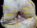
Additional images
-
 Capsule of right knee-joint (distended). Posterior aspect.
 -
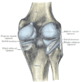
 Anterior view of knee.


External links
- The KNEEguru
- lljoints at The Anatomy Lesson by Wesley Norman (Georgetown University) (antkneejointopenflexed)
- Fibular collateral ligament injury explained
![]() This article incorporates text in the public domain from page 341 of the 20th edition of Gray's Anatomy (1918)
This article incorporates text in the public domain from page 341 of the 20th edition of Gray's Anatomy (1918)
This ligament-related article is a stub. You can help Wikipedia by expanding it. |