Echinocyte


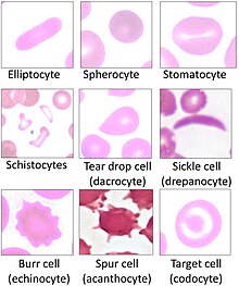
Echinocyte (from the Greek word echinos, meaning 'hedgehog' or 'sea urchin'), in human biology and medicine, refers to a form of red blood cell that has an abnormal cell membrane characterized by many small, evenly spaced thorny projections.[1][2] A more common term for these cells is burr cells.
Physiology
[edit]Echinocytes are frequently confused with acanthocytes, but the mechanism of cell membrane alteration is different. Echinocytosis is a reversible condition of red blood cells that is often merely an artifact produced by EDTA, which is used as an anticoagulant in sampled blood.[3] Echinocytes can be distinguished from acanthocytes by the shape of the projections, which are smaller and more numerous than in acanthocytes and are evenly spaced. Echinocytes also exhibit central pallor, or lightening of color in the center of the cell under Wright staining.[4]
Causes
[edit]In addition to appearing as an artifact of staining or drying, echinocytes are associated with:[5]
- Uremia and chronic kidney disease
- pyruvate kinase deficiency
- hypophosphatemia
- hyperlipidemia
- Phosphoglycerate kinase deficiency
- Disseminated malignancy
- Myeloproliferative disorders
- Vitamin E deficiency
- Early posttransfusion of RBC
Echinocytes, like acanthocytes, may be found in hyperlipidemia caused by liver dysfunction, but the lipids themselves do not integrate into the membrane. Instead, it is speculated that cell surface receptors on the red blood cells bind with HDL cholesterol, which induces the shape change.[6]
These cells were also shown to develop in vivo during hemodialysis, and disappear at the end of the procedure. The level of echinocytosis appeared to be related to the increase in blood viscosity that occurs during hemodialysis.[7]
The formation of echinocytes can also be induced by electric field pulses.[8] Alternating electric current produces modifications in the membranes of red blood cells, attributed to a higher permeability to water and a decreased tonicity, leading to the transformation into echinocytes.[9]
See also
[edit]References
[edit]- ^ Mentzer WC. Spiculated cells (echinocytes and acanthocytes) and target cells. UpToDate (release: 20.12- C21.4) [1]
- ^ Hoffman, R; Benz, EJ; Silberstein, LE; Heslop, H; Weitz J; Anastasi, J. (2012). Hematology: Basic Principles and Practice (6th ed.). Elsevier. ISBN 978-1-4377-2928-3.
- ^ MediaLab (July 12, 2013). "Burr Cells (Echinocytes)".
- ^ de Alarcon PA (Nov 30, 2011). "Acanthocytosis".
- ^ Tkachuk, Douglas C.; Hirschmann, Jan V., eds. (2007). Wintrobe's atlas of clinical hematology. Philadelphia, PA [etc.]: Lippincott Williams & Wilkins. ISBN 978-0781770231.
- ^ Owen, J S; Brown, D; Harry, D; McIntyre, N; Beaven, G; Isenberg, H; Gratzer, W (December 1985). "Erythrocyte echinocytosis in liver disease. Role of abnormal plasma high density lipoproteins". Journal of Clinical Investigation. 76 (6): 2275–85. doi:10.1172/JCI112237. PMC 424351. PMID 4077979.
- ^ Hasler, C R; Owen, G; Brunner, D; Reinhart, W (1998). "Echinocytosis induced by haemodialysis". Nephrology Dialysis Transplantation. 13 (12). Oxford Journals: 3132–3137. doi:10.1093/ndt/13.12.3132. PMID 9870478. Archived from the original on 2013-07-13.
- ^ Henszen, MM; Weske, M; Schwarz, S; Haest, CW; Deuticke, B (October–December 1997). "Electric field pulses induce reversible shape transformation of human erythrocytes". Mol Membr Biol. 14 (4): 195–204. doi:10.3109/09687689709048182. PMID 9491371.
- ^ Jeican, II; Matei, H; Istrate, A; Mironescu, E; Bâlici, S (April 2017). "Changes observed in erythrocyte cells exposed to an alternating current". Clujul Med. 90 (2): 154–60. doi:10.15386/cjmed-696. PMC 5433566. PMID 28559698.
