Galerina sulciceps
| Galerina sulciceps | |
|---|---|

| |
| Scientific classification | |
| Domain: | Eukaryota |
| Kingdom: | Fungi |
| Division: | Basidiomycota |
| Class: | Agaricomycetes |
| Order: | Agaricales |
| Family: | Hymenogastraceae |
| Genus: | Galerina |
| Species: | G. sulciceps
|
| Binomial name | |
| Galerina sulciceps | |

| |
| Natural distribution of Galerina sulciceps | |
| Synonyms[1][2] | |
|
Marasmius sulciceps Berk. (1847) | |
| Galerina sulciceps | |
|---|---|
| Gills on hymenium | |
| Cap is convex or flat | |
| Hymenium is adnate | |
| Stipe has a ring or is bare | |
| Spore print is yellow-orange to brown | |
| Ecology is saprotrophic | |
| Edibility is deadly | |
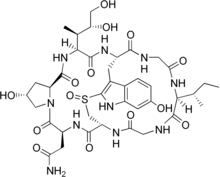
Galerina sulciceps is a dangerously toxic species of fungus in the family Strophariaceae, of the order Agaricales. It is distributed in tropical Indonesia and India, but has reportedly been found fruiting in European greenhouses on occasion. More toxic than the deathcap (Amanita phalloides), G. sulciceps has been shown to contain the toxins alpha- (
History and taxonomy
[edit]This species was first described in the literature as Marasmius sulciceps by English Naturalist Miles Joseph Berkeley in 1848, based on a specimen found four years earlier growing on old wood in Ceylon (modern-day Sri Lanka).[3] In 1898, Otto Kuntze transferred the species to Chamaeceras,[4] a genus that has since been subsumed back into Marasmius.[5] Because of its brown-colored spore print, Dutch mycologist Karel Bernard Boedijn transferred the species to the genus Phaeomarasmius 1938.[6] In 1951, he redescribed the species and transferred it to its current position in Galerina.[7] Rolf Singer's comprehensive taxonomical treatment of the Agaricales placed Galerina sulciceps in section Naucoriopsis of the genus Galerina, a subdivision first defined by French mycologist Robert Kühner in 1935.[8] This section includes small brown-spored fungi what when young have a cap margin that is curved inward, and thin-walled, obtuse, or acute-ended pleurocystidia that are not broadly rounded at the top.[9] All of the poisonous amatoxin-containing Galerina belong to section Naucoriopsis.[10]
Description
[edit]The cap is initially egg-shaped in young specimens, but changes shape as it matures, becoming convex and later more or less flat with a central depression. At the center of the cap is a roughly spherical umbo – a nipple-like protrusion. The cap is hygrophanous, meaning it changes color depending on its state of hydration: the color is tawny in moist specimens, changing to ochre with dark brown edges when dried.[11] The cap diameter is typically 1.5 to 4 cm (0.6 to 1.6 in), with a surface that is smooth, and almost gelatinous in consistency. The edge of the cap is thin and wavy, and is often split open. The gills are broadly adnate (broadly attached to the stalk slightly above the bottom of the gill, with most of the gill fused to the stem) to slightly decurrent (running down the length of the stem). Interspersed between the gills are shorter gills, called lamellulae, that start from the cap but do not reach the stem. The gills are broad (up to 4 mm) and thick at the base (1 mm), and when mature can develop veins that run between the gills on the undersurface of the cap. The stem is 0.4 to 2.5 cm (0.2 to 1.0 in) long, 0.15 to 0.3 cm (0.06 to 0.12 in) thick, and usually attached centrally to the underside of the cap, although it may sometimes be slightly off-center. Stems are solid, cylindrical, and may be pruinose (dusted with a very fine layer of powder).[1]
Berkeley's original description noted a resemblance to a small Marasmius peronatus,[3] a mushroom today known as Gymnopus peronatus.[12]
Microscopic characteristics
[edit]The spores are ellipsoid to almond-shaped, with dimensions of 7.2–9.7 by 4.5–5.8
Biochemistry
[edit]
Galerina sulciceps is deadly poisonous; one author opines it to be "perhaps the most toxic mushroom known to man",[13] while later studies of toxin concentrations in amanitin-containing mushrooms corroborate this view.[10][14] The symptoms of poisoning attributed to the mushroom have been noted to be relatively unusual: a local anesthesic effect, "pins and needles" sensation, and nausea without vomiting.[1] Although these clinical symptoms are inconsistent with those of amatoxin poisoning, the presence of
Habitat and distribution
[edit]This species grows on dead wood in tropical locales like Indonesia (Java and Sumatra), and near India (Sri Lanka), where it is prolific in some areas.[1] It is not found in North America.[20] In Germany, it has been found growing in greenhouses, and is known in the vernacular as the Gewächshaus-Häubling, meaning "greenhouse Galerina".[21] In one instance, the mushroom was discovered fruiting in dense groups in pots of orchids standing on moist conifer sawdust.[11]
See also
[edit]References
[edit]- ^ a b c d e f g Smith AH, Singer R (1963). A Monograph of the Genus Galerina Earle. New York, New York: Hafner Publishing. pp. 285–6.
- ^ "Chamaeceras sulciceps (Berk.) Kuntze 1898". MycoBank. International Mycological Association. Retrieved 2010-04-08.
- ^ a b Berkeley MJ. (1847). "Decades of fungi. Decade XV-XIX. Ceylon fungi". Journal of Botany, British and Foreign. 6: 479–514.
- ^ Kuntze O. (1898). Revisio generum plantarum (in Latin and German). Vol. 3. Leipzig, Germany: A. Felix. p. 457.
- ^ Kirk PM, Cannon PF, Minter DW, Stalpers JA (2008). Dictionary of the Fungi (10th ed.). Wallingford, UK: CAB International. p. 134. ISBN 978-0-85199-826-8.
- ^ a b Boedijn KB. (1938). "A poisonous species of the genus Phaeomarasmius". Extrait du Bulletin du Jardin botanique du Buitenzorg. Series 3. 16: 76–82.
- ^ Boedijn KB. (1951). "Some mycological notes". Sydowia. 5 (3–6): 211–9.
- ^ Kühner R. (1935). "Le Genre Galera (Fr.) Quélet" [The Genus Galera (Fr.) Quélet]. Encyclopédie Mycologique (in French). 7: 1–240.
- ^ Singer R. (1986). The Agaricales in Modern Taxonomy. Königstein im Taunus, Germany: Koeltz Scientific Books. pp. 673–4. ISBN 3-87429-254-1.
- ^ a b Enjalbert F, Cassanas G, Rapior S, Renault C, Chaumont J-P (2004). "Amatoxins in wood-rotting Galerina marginata". Mycologia. 96 (4): 720–9. doi:10.2307/3762106. JSTOR 3762106. PMID 21148893.
- ^ a b c Bresinsky A, Besl H (1989). A Colour Atlas of Poisonous Fungi: A Handbook for Pharmacists, Doctors, and Biologists. London, UK: Manson Publishing. pp. 40–1. ISBN 0-7234-1576-5.
- ^ "Names Record–Marasmius peronatus (Bolton) Fr". Index Fungorum. CAB International. Retrieved 2010-04-08.
- ^ Ammirati JF, Traquair JA, Horgen PA (1986). Poisonous Mushroom of Canada. Minneapolis, Minnesota: University of Minnesota Press. p. 81. ISBN 978-0-8166-1407-3.
- ^ Klán J. (1993). "Přehleb hub obsahujicich Amanitiny i Faloidiny" [The survey of fungi containing amanitins and phalloidins]. Časopis Lékařů Českých (in Czech). 132: 449–51.
- ^ Besl H. (1981). "Amatoxine im Gewächshaus: Galerina sulciceps, ein tropischer Giftpilz" [Amatoxins in greenhouses: Galerina sulciceps, a tropical poisonous mushroom]. Zeitschrift für Mykologie (in German). 47: 253–6.
- ^ Besl H, Mack P, Schmid-Heckel H (1984). "Giftpilze in den Gattungen Galerina und Lepiota" [Poisonous mushrooms in the genera Galerina and Lepiota]. Zeitschrift für Mykologie (in German). 50: 183–93.
- ^ Enjalbert F, Rapior S, Nouguier-Soulé J, Guillon S, Amouroux N, Cabot C (2002). "Treatment of amatoxin poisoning: 20-year retrospective analysis". Journal of Toxicology. Clinical Toxicology. 40 (6): 715–57. doi:10.1081/CLT-120014646. PMID 12475187. S2CID 22919515.
- ^ Chapuis J-R. (1981). "Bericht des Verbandstoxikologen fur das Jahr 1981" [Report by the toxicologist for 1981]. Schweizerische Zeitschrift für Pilzkunde (in German and French). 60 (9/10): 176–85.
- ^ Hall IR. (2003). Edible and Poisonous Mushrooms of the World. Portland, Oregon: Timber Press. p. 107. ISBN 0-88192-586-1.
- ^ Beuchat LR. (1987). Food and Beverage Mycology. Minneapolis, Minnesota: Springer. pp. 424–5. ISBN 978-0-442-21084-7.
- ^ Rebmann R. (2007). "Galerina sulciceps". Retrieved 2011-12-16.
