Medical ultrasonography
This does not have any sources. |


Diagnostic ultrasonography is the use of ultrasound to visualize hidden body structures. This medical imaging technique is used to display muscles, tendons, and other internal organs. It makes it easier to assess damage done to these structures.
Diagnostic sonography has a wide field of use, including emergency medicine, where it is regularly performed on patients which are in a state of shock or trauma. This test is known as Focused Assessment with Sonography for Trauma. It can detect any fluid in the body cavities. Doctors can then decide whether life-saving surgery must be performed.
Obstetric sonography
[change | change source]Is the name for a number of tests done during the pregnancy of a woman.
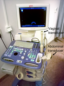
Gynecologic ultrasonography or gynecologic sonography refers to the application of medical ultrasonography to the female pelvic organs (specifically the uterus, the ovaries, and the fallopian tubes) as well as the bladder, the adnexa, and the recto-uterine pouch.
Gynecologic sonography is used often and throughout many different tests:
- to assess pelvic organs,
- to diagnose acute appendicitis[1]
- to diagnose and manage gynecologic problems including endometriosis, leiomyoma, adenomyosis, ovarian cysts and lesions,
- to identify adnexal masses, including ectopic pregnancy,
- to diagnose gynecologic cancer
- in infertility treatments to track the response of ovarian follicles to fertility medication (such as Pergonal). However, it often underestimates the true ovarian volume.[2]
References
[change | change source]- ↑ Caspi, B.; Zbar, AP.; Mavor, E.; Hagay, Z.; Appelman, Z. (Mar 2003). "The contribution of transvaginal ultrasound in the diagnosis of acute appendicitis: an observational study". Ultrasound Obstet Gynecol. 21 (3): 273–6. doi:10.1002/uog.72. PMID 12666223. S2CID 39843514.
- ↑ Rosendahl M, Ernst E, Rasmussen PE, Yding Andersen C (December 2008). "True ovarian volume is underestimated by two-dimensional transvaginal ultrasound measurement". Fertil. Steril. 93 (3): 995–998. doi:10.1016/j.fertnstert.2008.10.055. PMID 19108822.
