(Translated by https://www.hiragana.jp/)
Cells | March 2019 - Browse Articles
Next Issue
Volume 8, April
Previous Issue
Volume 8, February


Cells, Volume 8, Issue 3 (March 2019) – 87 articles

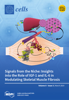
Cover Story (view full-size image): Fibrotic tissue deposition is generally associated with an altered interplay among the different cell components in muscle tissue, affecting regenerative processes. The expression of soluble mediators, including cytokines and growth factors, can modulate cell–cell communication, regulating critical steps of muscle healing, including regenerative fibrogenesis and myogenesis. In particular, IGF-1 and physiological levels of IL-6 play important roles in muscle regeneration, exerting a pro-myogenic activity, whereas elevated levels of IL-6 negatively impact muscle repair. Here, we focus on cellular and molecular mediators of skeletal muscle fibrosis and discuss how the altered expression of specific niche factors can impinge on the homeostatic mechanisms of tissue repair. View this paper.
  • Issues are regarded as officially published after their release is announced to the table of contents alert mailing list.
  • You may sign up for e-mail alerts to receive table of contents of newly released issues.
  • PDF is the official format for papers published in both, html and pdf forms. To view the papers in pdf format, click on the "PDF Full-text" link, and use the free Adobe Reader to open them.
Order results
Result details
Section
Select all
Export citation of selected articles as:
27 pages, 4449 KiB  
Review
Import of Non-Coding RNAs into Human Mitochondria: A Critical Review and Emerging Approaches
by Damien Jeandard, Anna Smirnova, Ivan Tarassov, Eric Barrey, Alexandre Smirnov and Nina Entelis
Cells 2019, 8(3), 286; https://doi.org/10.3390/cells8030286 - 26 Mar 2019
Cited by 62 | Viewed by 9699
Abstract
Mitochondria harbor their own genetic system, yet critically depend on the import of a number of nuclear-encoded macromolecules to ensure their expression. In all eukaryotes, selected non-coding RNAs produced from the nuclear genome are partially redirected into the mitochondria, where they participate in [...] Read more.
Mitochondria harbor their own genetic system, yet critically depend on the import of a number of nuclear-encoded macromolecules to ensure their expression. In all eukaryotes, selected non-coding RNAs produced from the nuclear genome are partially redirected into the mitochondria, where they participate in gene expression. Therefore, the mitochondrial RNome represents an intricate mixture of the intrinsic transcriptome and the extrinsic RNA importome. In this review, we summarize and critically analyze data on the nuclear-encoded transcripts detected in human mitochondria and outline the proposed molecular mechanisms of their mitochondrial import. Special attention is given to the various experimental approaches used to study the mitochondrial RNome, including some recently developed genome-wide and in situ techniques. Full article
(This article belongs to the Section Mitochondria)
Show Figures

Figure 1

15 pages, 4006 KiB  
Article
IL-6, IL-12, and IL-23 STAT-Pathway Genetic Risk and Responsiveness of Lymphocytes in Patients with Multiple Sclerosis
by Marina R. von Essen, Helle B. Søndergaard, Eva R.S. Petersen and Finn Sellebjerg
Cells 2019, 8(3), 285; https://doi.org/10.3390/cells8030285 - 26 Mar 2019
Cited by 23 | Viewed by 4767
Abstract
Multiple sclerosis (MS) is an immune-mediated demyelinating disease characterized by central nervous system (CNS) lymphocyte infiltration, abundant production of pro-inflammatory cytokines, and inappropriate activation of Th1 and Th17 cells, B cells, and innate immune cells. The etiology of MS is complex, and genetic [...] Read more.
Multiple sclerosis (MS) is an immune-mediated demyelinating disease characterized by central nervous system (CNS) lymphocyte infiltration, abundant production of pro-inflammatory cytokines, and inappropriate activation of Th1 and Th17 cells, B cells, and innate immune cells. The etiology of MS is complex, and genetic factors contribute to disease susceptibility. Genome-wide association studies (GWAS) have revealed numerous MS-risk alleles in the IL-6/STAT3, IL-12/STAT4, and IL-23/STAT3-pathways implicated in the differentiation of Th1 and Th17 cells. In this study, we investigated the signaling properties of these pathways in T, B, and NK cells from patients with relapsing-remitting MS (RRMS) and healthy controls, and assessed the genetic contribution to the activity of the pathways. This revealed a great variability in the level of STAT-pathway molecules and STAT activation between the cell types investigated. We also found a strong donor variation in IL-6, IL-12, and IL-23 responsiveness of primed CD4+ T cells. This variation could not be explained by a single MS-risk variant in a pathway component, or by an accumulation of multiple STAT-pathway MS-risk SNPs. The data of this study suggests that other factors in cohesion with the genetic background contribute to the responsiveness of the IL-6/STAT3, IL-12/STAT4, and IL-23/STAT3-pathways. Full article
(This article belongs to the Special Issue The Molecular and Cellular Basis for Multiple Sclerosis)
Show Figures

Figure 1

4 pages, 200 KiB  
Editorial
Emerging Cellular Therapies: T Cells and Beyond
by Stephen Todryk, Agnieszka Jozwik, Julian de Havilland and Joanna Hester
Cells 2019, 8(3), 284; https://doi.org/10.3390/cells8030284 - 26 Mar 2019
Cited by 7 | Viewed by 3912
Abstract
Cellular therapies, including those based on T cells, are becoming approved options for clinicians treating a range of diseases. Cytotoxic T lymphocytes (CTLs) can be modified ex vivo to express receptors such as chimeric antigen receptors (CARs) or T cell receptors, allowing them [...] Read more.
Cellular therapies, including those based on T cells, are becoming approved options for clinicians treating a range of diseases. Cytotoxic T lymphocytes (CTLs) can be modified ex vivo to express receptors such as chimeric antigen receptors (CARs) or T cell receptors, allowing them to target tumour cells when infused back into patients with particular cancers. CTLs specific for viruses can be purified ex vivo and reinfused into patients transplanted with haematopoietic stem cells to help combat viral reactivation. Regulatory T cells (Tregs) can be expanded ex vivo for infusion into patients with autoimmunity or allergy, or into those at risk of rejecting transplanted cells or tissues, or suffering graft versus host disease. Effector and regulatory T cells can also be generated by infusion of patient-derived dendritic cells (DCs) conditioned in ways to elicit anti-tumour immunity (CTLs) or Tregs. All such therapies are resource-heavy (particularly in process regulation) and so must be initially targeted to patients that have limited treatment options, but also where they have a chance of being effective. Full article
(This article belongs to the Special Issue Emerging Cellular Therapies: T Cells and Beyond)
19 pages, 9267 KiB  
Article
Ethanol-Induced Mitochondrial Damage in Sertoli Cells is Associated with Parkin Overexpression and Activation of Mitophagy
by Nabil Eid, Yuko Ito, Akio Horibe, Yoshinori Otsuki and Yoichi Kondo
Cells 2019, 8(3), 283; https://doi.org/10.3390/cells8030283 - 25 Mar 2019
Cited by 20 | Viewed by 6392
Abstract
This study was conducted to elucidate the involvement of the PINK1-Parkin pathway in ethanol-induced mitophagy among Sertoli cells (SCs). In the research, adult rats were given intraperitoneal injections of ethanol (5 gm/kg) and sacrificed at various time periods within 24 h. Transmission electron [...] Read more.
This study was conducted to elucidate the involvement of the PINK1-Parkin pathway in ethanol-induced mitophagy among Sertoli cells (SCs). In the research, adult rats were given intraperitoneal injections of ethanol (5 gm/kg) and sacrificed at various time periods within 24 h. Transmission electron microscopy was applied to reveal enhanced mitochondrial damage in SCs of the ethanol-treated rats (ETRs) in association with a significant increase in numbers of mitophagic vacuoles (mitophagosomes and autolysosomes) in contrast to very low levels in a control group treated with phosphate-buffered saline (PBS). This enhancement was ultra-structurally verified via observation of trapped mitochondria within LC3-labeled membranes, upregulation of LC3 protein levels, colocalization of LC3 and cytochrome c, and reduced expression of mitochondrial proteins. Importantly, Parkin expression was found to be upregulated in ETR SCs, specifically in mitochondria and mitophagosomes in addition to colocalization with PINK1 and pan-cathepsin, indicating augmented mitophagy. Transcription factor EB (TFEB, a transcription factor for autophagy and mitophagy proteins) was also found to be upregulated in nuclei of ETR SCs and associated with enhanced expression of iNOS. Enhanced Parkin-related mitophagy in ETR SCs may be a protective mechanism with therapeutic implications. To the authors’ knowledge, this is the first report demonstrating the ultrastructural characteristics and molecular mechanisms of Parkin-related mitophagy in ETR SCs. Full article
(This article belongs to the Special Issue Autophagy in Tissue Injury and Homeostasis)
Show Figures

Figure 1

20 pages, 32131 KiB  
Article
Characteristics and Potentiality of Human Adipose-Derived Stem Cells (hASCs) Obtained from Enzymatic Digestion of Fat Graft
by Pietro Gentile, Maria Serena Piccinno and Claudio Calabrese
Cells 2019, 8(3), 282; https://doi.org/10.3390/cells8030282 - 25 Mar 2019
Cited by 70 | Viewed by 5854
Abstract
Human adipose-derived stem cells localize in the stromal-vascular portion, and can be ex vivo isolated using a combination of washing steps and enzymatic digestion. For this study, we undertook a histological evaluation of traditional fat graft compared with fat graft enriched with stromal [...] Read more.
Human adipose-derived stem cells localize in the stromal-vascular portion, and can be ex vivo isolated using a combination of washing steps and enzymatic digestion. For this study, we undertook a histological evaluation of traditional fat graft compared with fat graft enriched with stromal vascular fraction cells isolated by the Celution™ system to assess the interactions between cells and adipose tissue before the breast injection. In addition, we reported on histological analyses of biopsies derived from fat grafted (traditional or enriched with SVFs) in the breast in order to assess the quality of the adipose tissue, fibrosis and vessels. The hASCs derived from enzymatic digestion were systematically characterized for growth features, phenotype and multi-potent differentiation potential. They fulfill the definition of mesenchymal stem cells, albeit with a higher neural phenotype profile. These cells also express genes that constitute the core circuitry of self-renewal such as OCT4, SOX2, NANOG and neurogenic lineage genes such as NEUROD1, PAX6 and SOX3. Such findings support the hypothesis that hASCs may have a potential usefulness in neurodegenerative conditions. These data can be helpful for the development of new therapeutic approaches in personalized medicine to assess safety and efficacy of the breast reconstruction. Full article
(This article belongs to the Special Issue Stem Cells in Personalized Medicine)
Show Figures

Graphical abstract

15 pages, 2863 KiB  
Article
Novel Flavivirus Antiviral That Targets the Host Nuclear Transport Importin αあるふぁ/βべーた1 Heterodimer
by Sundy N. Y. Yang, Sarah C. Atkinson, Johanna E. Fraser, Chunxiao Wang, Belinda Maher, Noelia Roman, Jade K. Forwood, Kylie M. Wagstaff, Natalie A. Borg and David A. Jans
Cells 2019, 8(3), 281; https://doi.org/10.3390/cells8030281 - 24 Mar 2019
Cited by 33 | Viewed by 7736
Abstract
Dengue virus (DENV) threatens almost 70% of the world’s population, with no effective vaccine or therapeutic currently available. A key contributor to infection is nuclear localisation in the infected cell of DENV nonstructural protein 5 (NS5) through the action of the host importin [...] Read more.
Dengue virus (DENV) threatens almost 70% of the world’s population, with no effective vaccine or therapeutic currently available. A key contributor to infection is nuclear localisation in the infected cell of DENV nonstructural protein 5 (NS5) through the action of the host importin (IMP) αあるふぁ/βべーた1 proteins. Here, we used a range of microscopic, virological and biochemical/biophysical approaches to show for the first time that the small molecule GW5074 has anti-DENV action through its novel ability to inhibit NS5–IMPαあるふぁ/βべーた1 interaction in vitro as well as NS5 nuclear localisation in infected cells. Strikingly, GW5074 not only inhibits IMPαあるふぁ binding to IMPβべーた1, but can dissociate preformed IMPαあるふぁ/βべーた1 heterodimer, through targeting the IMPαあるふぁ armadillo (ARM) repeat domain to impact IMPαあるふぁ thermal stability and αあるふぁ-helicity, as shown using analytical ultracentrifugation, thermostability analysis and circular dichroism measurements. Importantly, GW5074 has strong antiviral activity at low µM concentrations against not only DENV-2, but also zika virus and West Nile virus. This work highlights DENV NS5 nuclear targeting as a viable target for anti-flaviviral therapeutics. Full article
(This article belongs to the Special Issue Nuclear Transport in Ageing and Diseases)
Show Figures

Figure 1

36 pages, 7988 KiB  
Review
2,4 Dinitrophenol as Medicine
by John G. Geisler
Cells 2019, 8(3), 280; https://doi.org/10.3390/cells8030280 - 23 Mar 2019
Cited by 76 | Viewed by 27849
Abstract
In the sanctity of pure drug discovery, objective reasoning can become clouded when pursuing ideas that appear unorthodox, but are spot on physiologically. To put this into historical perspective, it was an unorthodox idea in the 1950’s to suggest that warfarin, a rat [...] Read more.
In the sanctity of pure drug discovery, objective reasoning can become clouded when pursuing ideas that appear unorthodox, but are spot on physiologically. To put this into historical perspective, it was an unorthodox idea in the 1950’s to suggest that warfarin, a rat poison, could be repositioned into a breakthrough drug in humans to protect against strokes as a blood thinner. Yet it was approved in 1954 as Coumadin® and has been prescribed to billions of patients as a standard of care. Similarly, no one can forget the horrific effects of thalidomide, prescribed or available without a prescription, as both a sleeping pill and “morning sickness” anti-nausea medication targeting pregnant women in the 1950’s. The “thalidomide babies” became the case-in-point for the need of strict guidelines by the U.S. Food & Drug Administration (FDA) or full multi-species teratogenicity testing before drug approval. More recently it was found that thalidomide is useful in graft versus host disease, leprosy and resistant tuberculosis treatment, and as an anti-angiogenesis agent as a breakthrough drug for multiple myeloma (except for pregnant female patients). Decades of diabetes drug discovery research has historically focused on every possible angle, except, the energy-out side of the equation, namely, raising mitochondrial energy expenditure with chemical uncouplers. The idea of “social responsibility” allowed energy-in agents to be explored and the portfolio is robust with medicines of insulin sensitizers, insulin analogues, secretagogues, SGLT2 inhibitors, etc., but not energy-out medicines. The primary reason? It appeared unorthodox, to return to exploring a drug platform used in the 1930s in over 100,000 obese patients used for weight loss. This is over 80-years ago and prior to Dr Peter Mitchell explaining the mechanism of how mitochondrial uncouplers, like 2,4-dinitrophenol (DNP) even worked by three decades later in 1961. Although there is a clear application for metabolic disease, it was not until recently that this platform was explored for its merit at very low, weight-neutral doses, for treating insidious human illnesses and completely unrelated to weight reduction. It is known that mitochondrial uncouplers specifically target the entire organelle’s physiology non-genomically. It has been known for years that many neuromuscular and neurodegenerative diseases are associated with overt production of reactive oxygen species (ROSs), a rise in isoprostanes (biomarker of mitochondrial ROSs in urine or blood) and poor calcium (Ca2+) handing. It has also been known that mitochondrial uncouplers lower ROS production and Ca2+ overload. There is evidence that elevation of isoprostanes precedes disease onset, in Alzheimer’s Disease (AD). It is also curious, why so many neurodegenerative diseases of known and unknown etiology start at mid-life or later, such as Multiple Sclerosis (MS), Huntington Disease (HD), AD, Parkinson Disease, and Amyotrophic Lateral Sclerosis (ALS). Is there a relationship to a buildup of mutations that are sequestered over time due to ROSs exceeding the rate of repair? If ROS production were managed, could disease onset due to aging be delayed or prevented? Is it possible that most, if not all neurodegenerative diseases are manifested through mitochondrial dysfunction? Although DNP, a historic mitochondrial uncoupler, was used in the 1930s at high doses for obesity in well over 100,000 humans, and so far, it has never been an FDA-approved drug. This review will focus on the application of using DNP, but now, repositioned as a potential disease-modifying drug for a legion of insidious diseases at much lower and paradoxically, weight neutral doses. DNP will be addressed as a treatment for “metabesity”, an emerging term related to the global comorbidities associated with the over-nutritional phenotype; obesity, diabetes, nonalcoholic steatohepatitis (NASH), metabolic syndrome, cardiovascular disease, but including neurodegenerative disorders and accelerated aging. Some unexpected drug findings will be discussed, such as DNP’s induction of neurotrophic growth factors involved in neuronal heath, learning and cognition. For the first time in 80’s years, the FDA has granted (to Mitochon Pharmaceutical, Inc., Blue Bell, PA, USA) an open Investigational New Drug (IND) approval to begin rigorous clinical testing of DNP for safety and tolerability, including for the first ever, pharmacokinetic profiling in humans. Successful completion of Phase I clinical trial will open the door to explore the merits of DNP as a possible treatment of people with many truly unmet medical needs, including those suffering from HD, MS, PD, AD, ALS, Duchenne Muscular Dystrophy (DMD), and Traumatic Brain Injury (TBI). Full article
(This article belongs to the Section Intracellular and Plasma Membranes)
Show Figures

Figure 1

13 pages, 2493 KiB  
Article
FM19G11-Loaded Gold Nanoparticles Enhance the Proliferation and Self-Renewal of Ependymal Stem Progenitor Cells Derived from ALS Mice
by Stefania Marcuzzo, Davide Isaia, Silvia Bonanno, Claudia Malacarne, Paola Cavalcante, Antonella Zacheo, Valentino Laquintana, Nunzio Denora, Barbara Sanavio, Elisa Salvati, Patrizia Andreozzi, Francesco Stellacci, Silke Krol, Maravillas Mellado-López, Renato Mantegazza, Victoria Moreno-Manzano and Pia Bernasconi
Cells 2019, 8(3), 279; https://doi.org/10.3390/cells8030279 - 23 Mar 2019
Cited by 27 | Viewed by 5132
Abstract
Amyotrophic lateral sclerosis (ALS) is a progressive neurodegenerative disease affecting motor neurons. In ALS mice, neurodegeneration is associated with the proliferative restorative attempts of ependymal stem progenitor cells (epSPCs) that normally lie in a quiescent in the spinal cord. Thus, modulation of the [...] Read more.
Amyotrophic lateral sclerosis (ALS) is a progressive neurodegenerative disease affecting motor neurons. In ALS mice, neurodegeneration is associated with the proliferative restorative attempts of ependymal stem progenitor cells (epSPCs) that normally lie in a quiescent in the spinal cord. Thus, modulation of the proliferation of epSPCs may represent a potential strategy to counteract neurodegeneration. Recent studies demonstrated that FM19G11, a hypoxia-inducible factor modulator, induces epSPC self-renewal and proliferation. The aim of the study was to investigate whether FM19G11-loaded gold nanoparticles (NPs) can affect self-renewal and proliferation processes in epSPCs isolated from G93A-SOD1 mice at disease onset. We discovered elevated levels of SOX2, OCT4, AKT1, and AKT3, key genes associated with pluripotency, self-renewal, and proliferation, in G93A-SOD1 epSPCs at the transcriptional and protein levels after treatment with FM19G11-loaded NPs. We also observed an increase in the levels of the mitochondrial uncoupling protein (UCP) gene in treated cells. FM19G11-loaded NPs treatment also affected the expression of the cell cycle-related microRNA (miR)-19a, along with its target gene PTEN, in G93A-SOD1 epSPCs. Overall our findings establish the significant impact of FM19G11-loaded NPs on the cellular pathways involved in self-renewal and proliferation in G93A-SOD1 epSPCs, thus providing an impetus to the design of novel tailored approaches to delay ALS disease progression. Full article
(This article belongs to the Special Issue Stem Cells and Degenerative Diseases)
Show Figures

Figure 1

15 pages, 1100 KiB  
Review
Recent Progress on the Localization of the Spindle Assembly Checkpoint Machinery to Kinetochores
by Zhen Dou, Diogjena Katerina Prifti, Ping Gui, Xing Liu, Sabine Elowe and Xuebiao Yao
Cells 2019, 8(3), 278; https://doi.org/10.3390/cells8030278 - 23 Mar 2019
Cited by 30 | Viewed by 9805
Abstract
Faithful chromosome segregation during mitosis is crucial for maintaining genome stability. The spindle assembly checkpoint (SAC) is a surveillance mechanism that ensures accurate mitotic progression. Defective SAC signaling leads to premature sister chromatid separation and aneuploid daughter cells. Mechanistically, the SAC couples the [...] Read more.
Faithful chromosome segregation during mitosis is crucial for maintaining genome stability. The spindle assembly checkpoint (SAC) is a surveillance mechanism that ensures accurate mitotic progression. Defective SAC signaling leads to premature sister chromatid separation and aneuploid daughter cells. Mechanistically, the SAC couples the kinetochore microtubule attachment status to the cell cycle progression machinery. In the presence of abnormal kinetochore microtubule attachments, the SAC prevents the metaphase-to-anaphase transition through a complex kinase-phosphatase signaling cascade which results in the correct balance of SAC components recruited to the kinetochore. The correct kinetochore localization of SAC proteins is a prerequisite for robust SAC signaling and, hence, accurate chromosome segregation. Here, we review recent progresses on the kinetochore recruitment of core SAC factors. Full article
(This article belongs to the Special Issue Molecular Regulation of Mitosis and Its Role in Disease)
Show Figures

Figure 1

16 pages, 4858 KiB  
Article
A New Targeted Lipidomics Approach Reveals Lipid Droplets in Liver, Muscle and Heart as a Repository for Diacylglycerol and Ceramide Species in Non-Alcoholic Fatty Liver
by Christina Preuss, Tomas Jelenik, Kálmán Bódis, Karsten Müssig, Volker Burkart, Julia Szendroedi, Michael Roden and Daniel F. Markgraf
Cells 2019, 8(3), 277; https://doi.org/10.3390/cells8030277 - 22 Mar 2019
Cited by 46 | Viewed by 7710
Abstract
Obesity is frequently associated with excessive accumulation of lipids in ectopic tissue and presents a major risk factor for type 2 diabetes (T2D) and non-alcoholic fatty liver disease (NAFLD). Diacylglycerols (DAGs) and ceramides (CERs) were identified as key players in lipid-induced insulin resistance, [...] Read more.
Obesity is frequently associated with excessive accumulation of lipids in ectopic tissue and presents a major risk factor for type 2 diabetes (T2D) and non-alcoholic fatty liver disease (NAFLD). Diacylglycerols (DAGs) and ceramides (CERs) were identified as key players in lipid-induced insulin resistance, typical for such diseases. Recent results suggest that the subcellular distribution of these lipids affects their lipotoxic properties. However, the subcellular dynamics of these lipids and the role of lipid droplets (LDs) as a potential storage site is not understood. Here, we developed a liquid chromatography triple quadrupole mass spectrometry (LC-MS/MS)-method for the rapid and simultaneous quantification of DAG and CER species in tissue sample fractions. The assay is characterized by excellent recovery of analytes, limit of quantification, accuracy and precision. We established a fractionation protocol that allows the separation of subcellular tissue fractions. This method was subsequently tested to measure the concentration of DAGs and CERs in subcellular fractions of human muscle and several mouse tissues. In a mouse model of NAFLD, application of this method revealed a prominent role for LDs as repository for lipotoxic DAG and CER species. In conclusion, the new method proved as a valuable tool to analyse the subcellular dynamics of lipotoxins, related to the pathogenesis of insulin resistance, T2D and NAFLD. Full article
(This article belongs to the Section Intracellular and Plasma Membranes)
Show Figures

Figure 1

20 pages, 4859 KiB  
Article
CD44s Assembles Hyaluronan Coat on Filopodia and Extracellular Vesicles and Induces Tumorigenicity of MKN74 Gastric Carcinoma Cells
by Kai Härkönen, Sanna Oikari, Heikki Kyykallio, Janne Capra, Sini Hakkola, Kirsi Ketola, Uma Thanigai Arasu, George Daaboul, Andrew Malloy, Carla Oliveira, Otto Jokelainen, Reijo Sironen, Jaana M. Hartikainen and Kirsi Rilla
Cells 2019, 8(3), 276; https://doi.org/10.3390/cells8030276 - 22 Mar 2019
Cited by 31 | Viewed by 6237
Abstract
CD44 is a multifunctional adhesion molecule typically upregulated in malignant, inflamed and injured tissues. Due to its ability to bind multiple ligands present in the tumor microenvironment, it promotes multiple cellular functions related to tumorigenesis. Recent data has shown that CD44 and its [...] Read more.
CD44 is a multifunctional adhesion molecule typically upregulated in malignant, inflamed and injured tissues. Due to its ability to bind multiple ligands present in the tumor microenvironment, it promotes multiple cellular functions related to tumorigenesis. Recent data has shown that CD44 and its principal ligand hyaluronan (HA) are carried by extracellular vesicles (EV) derived from stem and tumor cells, but the role of CD44 in EV shedding has not been studied so far. To answer this question, we utilized CD44-negative human gastric carcinoma cell line MKN74 manipulated to stably express CD44 standard form (CD44s). The effect of CD44s expression on HA metabolism, EV secretion, morphology and growth of these cells was studied. Interestingly, HAS2 and HYAL2 expression levels were significantly upregulated in CD44s-expressing cells. Cell-associated HA levels were significantly increased, while HA levels in the culture medium of CD44s-positive cells was lower compared to CD44s-negative MOCK cells. CD44s expression had no significant effect on the proliferation capacity of cells, but cells showed diminished contact inhibition. Superresolution imaging revealed that CD44s and HA were accumulated on filopodia and EVs secreted from CD44s-positive cells, but no differences in total numbers of secreted EV between CD44s-negative and -positive cells was detected. In 3D cultures, CD44s-expressing cells had an enhanced invasion capacity in BME gel and increased spheroidal growth when cultured in collagen I gel. No significant differences in mitotic activity, tumor size or morphology were detected in CAM assays. However, a significant increase in HA staining coverage was detected in CD44s-positive tumors. Interestingly, CD44s-positive EVs embedded in HA-rich matrix were detected in the stromal areas of tumors. The results indicate that CD44s expression significantly increases the HA binding capacity of gastric cancer cells, while the secreted HA is downregulated. CD44s is also carried by EVs secreted by CD44s-expressing cells. These findings highlight the potential usefulness of CD44s and its ligands as multipurpose EV biomarkers, because they are upregulated in inflammatory, injured, and cancer cells and accumulate on the surface of EVs secreted in these situations. Full article
(This article belongs to the Special Issue Tumor Microenvironment: Interaction and Metabolism)
Show Figures

Figure 1

19 pages, 1199 KiB  
Review
Mitochondrial Retrograde Signalling and Metabolic Alterations in the Tumour Microenvironment
by Dongki Yang and Jaehong Kim
Cells 2019, 8(3), 275; https://doi.org/10.3390/cells8030275 - 22 Mar 2019
Cited by 41 | Viewed by 7143
Abstract
This review explores the molecular mechanisms that may be responsible for mitochondrial retrograde signalling related metabolic reprogramming in cancer and host cells in the tumour microenvironment and provides a summary of recent updates with regard to the functional modulation of diverse cells in [...] Read more.
This review explores the molecular mechanisms that may be responsible for mitochondrial retrograde signalling related metabolic reprogramming in cancer and host cells in the tumour microenvironment and provides a summary of recent updates with regard to the functional modulation of diverse cells in the tumour microenvironment. Full article
(This article belongs to the Special Issue Mitochondrial Metabolic Reprogramming and Nuclear Crosstalk in Cancer)
Show Figures

Figure 1

14 pages, 865 KiB  
Review
Strategies to Circumvent Host Innate Immune Response by Hepatitis C Virus
by Tapas Patra, Ratna B. Ray and Ranjit Ray
Cells 2019, 8(3), 274; https://doi.org/10.3390/cells8030274 - 22 Mar 2019
Cited by 13 | Viewed by 6349
Abstract
Innate immune responses generate interferons, proinflammatory cytokines, complement activation, and natural killer (NK) cell response. Ultimately, this leads to the induction of a robust virus-specific adaptive immunity. Although the host innate immune system senses and responds to eliminate virus infection, hepatitis C virus [...] Read more.
Innate immune responses generate interferons, proinflammatory cytokines, complement activation, and natural killer (NK) cell response. Ultimately, this leads to the induction of a robust virus-specific adaptive immunity. Although the host innate immune system senses and responds to eliminate virus infection, hepatitis C virus (HCV) evades immune attack and establishes persistent infection within the liver. Spontaneous clearance of HCV infection is associated with a prompt induction of innate immunity generated in an infected host. In this review, we have highlighted the current knowledge of our understanding of host–HCV interactions, especially for endogenous interferon production, proinflammatory response, NK cell response, and complement activation, which may impair the generation of a strong adaptive immune response for establishment of chronicity. The information may provide novel strategies in augmenting therapeutic intervention against HCV. Full article
(This article belongs to the Special Issue Hepatitis C Virus and Host Interactions)
Show Figures

Graphical abstract

21 pages, 3458 KiB  
Article
N-Glycomic and Transcriptomic Changes Associated with CDX1 mRNA Expression in Colorectal Cancer Cell Lines
by Stephanie Holst, Jennifer L. Wilding, Kamila Koprowska, Yoann Rombouts and Manfred Wuhrer
Cells 2019, 8(3), 273; https://doi.org/10.3390/cells8030273 - 22 Mar 2019
Cited by 20 | Viewed by 6588
Abstract
The caudal-related homeobox protein 1 (CDX1) is a transcription factor, which is important in the development, differentiation, and homeostasis of the gut. Although the involvement of CDX genes in the regulation of the expression levels of a few glycosyltransferases has been shown, associations [...] Read more.
The caudal-related homeobox protein 1 (CDX1) is a transcription factor, which is important in the development, differentiation, and homeostasis of the gut. Although the involvement of CDX genes in the regulation of the expression levels of a few glycosyltransferases has been shown, associations between glycosylation phenotypes and CDX1 mRNA expression have hitherto not been well studied. Triggered by our previous study, we here characterized the N-glycomic phenotype of 16 colon cancer cell lines, selected for their differential CDX1 mRNA expression levels. We found that high CDX1 mRNA expression associated with a higher degree of multi-fucosylation on N-glycans, which is in line with our previous results and was supported by up-regulated gene expression of fucosyltransferases involved in antenna fucosylation. Interestingly, hepatocyte nuclear factors (HNF)4A and HNF1A were, among others, positively associated with high CDX1 mRNA expression and have been previously proven to regulate antenna fucosylation. Besides fucosylation, we found that high CDX1 mRNA expression in cancer cell lines also associated with low levels of sialylation and galactosylation and high levels of bisection on N-glycans. Altogether, our data highlight a possible role of CDX1 in altering the N-glycosylation of colorectal cancer cells, which is a hallmark of tumor development. Full article
(This article belongs to the Special Issue Functional Glycomics)
Show Figures

Figure 1

17 pages, 887 KiB  
Review
C-Met as a Key Factor Responsible for Sustaining Undifferentiated Phenotype and Therapy Resistance in Renal Carcinomas
by Paulina Marona, Judyta Górka, Jerzy Kotlinowski, Marcin Majka, Jolanta Jura and Katarzyna Miekus
Cells 2019, 8(3), 272; https://doi.org/10.3390/cells8030272 - 22 Mar 2019
Cited by 27 | Viewed by 4886
Abstract
C-Met tyrosine kinase receptor plays an important role under normal and pathological conditions. In tumor cells’ overexpression or incorrect activation of c-Met, this leads to stimulation of proliferation, survival and increase of motile activity. This receptor is also described as a marker of [...] Read more.
C-Met tyrosine kinase receptor plays an important role under normal and pathological conditions. In tumor cells’ overexpression or incorrect activation of c-Met, this leads to stimulation of proliferation, survival and increase of motile activity. This receptor is also described as a marker of cancer initiating cells. The latest research shows that the c-Met receptor has an influence on the development of resistance to targeted cancer treatment. High c-Met expression and activation in renal cell carcinomas is associated with the progression of the disease and poor survival of patients. C-Met receptor has become a therapeutic target in kidney cancer. However, the therapies used so far using c-Met tyrosine kinase inhibitors demonstrate resistance to treatment. On the other hand, the c-Met pathway may act as an alternative target pathway in tumors that are resistant to other therapies. Combination treatment together with c-Met inhibitor reduces tumor growth, vascularization and pro-metastatic behavior and results in suppressed mesenchymal phenotype and vascular endothelial growth factor (VEGF) secretion. Recently, it has been shown that the acquirement of mesenchymal phenotype or lack of cell differentiation might be related to the presence of the c-Met receptor and is consequently responsible for therapy resistance. This review presents the results from recent studies identifying c-Met as an important factor in renal carcinomas being responsible for tumor growth, progression and metastasis, indicating the role of c-Met in resistance to antitumor therapy and demonstrating the pivotal role of c-Met in supporting mesenchymal cell phenotype. Full article
(This article belongs to the Special Issue Receptor Tyrosine Kinases in Health and Disease)
Show Figures

Graphical abstract

15 pages, 5098 KiB  
Review
Lamina Associated Domains and Gene Regulation in Development and Cancer
by Silke J. A. Lochs, Samy Kefalopoulou and Jop Kind
Cells 2019, 8(3), 271; https://doi.org/10.3390/cells8030271 - 21 Mar 2019
Cited by 44 | Viewed by 11525
Abstract
The nuclear lamina (NL) is a thin meshwork of filaments that lines the inner nuclear membrane, thereby providing a platform for chromatin binding and supporting genome organization. Genomic regions contacting the NL are lamina associated domains (LADs), which contain thousands of genes that [...] Read more.
The nuclear lamina (NL) is a thin meshwork of filaments that lines the inner nuclear membrane, thereby providing a platform for chromatin binding and supporting genome organization. Genomic regions contacting the NL are lamina associated domains (LADs), which contain thousands of genes that are lowly transcribed, and enriched for repressive histone modifications. LADs are dynamic structures that shift spatial positioning in accordance with cell-type specific gene expression changes during differentiation and development. Furthermore, recent studies have linked the disruption of LADs and alterations in the epigenome with the onset of diseases such as cancer. Here we focus on the role of LADs and the NL in gene regulation during development and cancer. Full article
(This article belongs to the Collection Lamins and Laminopathies)
Show Figures

Figure 1

20 pages, 2550 KiB  
Review
Two Birds, One Stone: Double Hits on Tumor Growth and Lymphangiogenesis by Targeting Vascular Endothelial Growth Factor Receptor 3
by Ming-Chuan Hsu, Mei-Ren Pan and Wen-Chun Hung
Cells 2019, 8(3), 270; https://doi.org/10.3390/cells8030270 - 21 Mar 2019
Cited by 47 | Viewed by 10480
Abstract
Vascular endothelial growth factor receptor 3 (VEGFR3) has been known for its involvement in tumor-associated lymphangiogenesis and lymphatic metastasis. The VEGFR3 signaling is stimulated by its main cognate ligand, vascular endothelial growth factor C (VEGF-C), which in turn promotes tumor progression. Activation of [...] Read more.
Vascular endothelial growth factor receptor 3 (VEGFR3) has been known for its involvement in tumor-associated lymphangiogenesis and lymphatic metastasis. The VEGFR3 signaling is stimulated by its main cognate ligand, vascular endothelial growth factor C (VEGF-C), which in turn promotes tumor progression. Activation of VEGF-C/VEGFR3 signaling in lymphatic endothelial cells (LECs) was shown to enhance the proliferation of LECs and the formation of lymphatic vessels, leading to increased lymphatic metastasis of tumor cells. In the past decade, the expression and pathological roles of VEGFR3 in tumor cells have been described. Moreover, the VEGF-C/VEGFR3 axis has been implicated in regulating immune tolerance and suppression. Therefore, the inhibition of the VEGF-C/VEGFR3 axis has emerged as an important therapeutic strategy for the treatment of cancer. In this review, we discuss the current findings related to VEGF-C/VEGFR3 signaling in cancer progression and recent advances in the development of therapeutic drugs targeting VEGF-C/VEGFR3. Full article
(This article belongs to the Special Issue Receptor Tyrosine Kinases in Health and Disease)
Show Figures

Figure 1

16 pages, 4678 KiB  
Article
Discovery of Small Molecules That Target Vascular Endothelial Growth Factor Receptor-2 Signalling Pathway Employing Molecular Modelling Studies
by Shailima Rampogu, Ayoung Baek, Chanin Park, Minky Son, Shraddha Parate, Saravanan Parameswaran, Yohan Park, Baji Shaik, Ju Hyun Kim, Seok Ju Park and Keun Woo Lee
Cells 2019, 8(3), 269; https://doi.org/10.3390/cells8030269 - 21 Mar 2019
Cited by 17 | Viewed by 4241
Abstract
Angiogenesis is defined as the formation of new blood vessels and is a key phenomenon manifested in a host of cancers during which tyrosine kinases play a crucial role. Vascular endothelial growth factor receptor-2 (VEGFR-2) is pivotal in cancer angiogenesis, which warrants the [...] Read more.
Angiogenesis is defined as the formation of new blood vessels and is a key phenomenon manifested in a host of cancers during which tyrosine kinases play a crucial role. Vascular endothelial growth factor receptor-2 (VEGFR-2) is pivotal in cancer angiogenesis, which warrants the urgency of discovering new anti-angiogenic inhibitors that target the signalling pathways. To obtain this objective, a structure-based pharmacophore model was built from the drug target VEGFR-2 (PDB code: 4AG8), complexed with axitinib and was subsequently validated and employed as a 3D query to retrieve the candidate compounds with the key inhibitory features. The model was escalated to molecular docking studies resulting in seven candidate compounds. The molecular docking studies revealed that the seven compounds displayed a higher dock score than the reference-cocrystallised compound. The GROningen MAchine for Chemical Simulations (GROMACS) package guided molecular dynamics (MD) results determined their binding mode and affirmed stable root mean square deviation. Furthermore, these compounds have preserved their key interactions with the residues Glu885, Glu917, Cys919 and Asp1046. The obtained findings deem that the seven compounds could act as novel anti-angiogenic inhibitors and may further assist as the prototype in designing and developing new inhibitors. Full article
(This article belongs to the Special Issue Protein Kinases: Signalling and Disease)
Show Figures

Graphical abstract

12 pages, 3836 KiB  
Article
Promethin Is a Conserved Seipin Partner Protein
by Inês G. Castro, Michal Eisenberg-Bord, Elisa Persiani, Justin J. Rochford, Maya Schuldiner and Maria Bohnert
Cells 2019, 8(3), 268; https://doi.org/10.3390/cells8030268 - 21 Mar 2019
Cited by 50 | Viewed by 7137
Abstract
Seipin (BSCL2/SPG17) is a key factor in lipid droplet (LD) biology, and its dysfunction results in severe pathologies, including the fat storage disease Berardinelli-Seip congenital lipodystrophy type 2, as well as several neurological seipinopathies. Despite its importance for human health, the molecular role [...] Read more.
Seipin (BSCL2/SPG17) is a key factor in lipid droplet (LD) biology, and its dysfunction results in severe pathologies, including the fat storage disease Berardinelli-Seip congenital lipodystrophy type 2, as well as several neurological seipinopathies. Despite its importance for human health, the molecular role of seipin is still enigmatic. Seipin is evolutionarily conserved from yeast to humans. In yeast, seipin was recently found to cooperate with the lipid droplet organization (LDO) proteins, Ldo16 and Ldo45, two structurally-related proteins involved in LD function and identity that display remote homology to the human protein promethin/TMEM159. In this study, we show that promethin is indeed an LD-associated protein that forms a complex with seipin, and its localization to the LD surface can be modulated by seipin expression levels. We thus identify promethin as a novel seipin partner protein. Full article
Show Figures

Graphical abstract

12 pages, 624 KiB  
Perspective
Why is NMNAT Protective against Neuronal Cell Death and Axon Degeneration, but Inhibitory of Axon Regeneration?
by Bor Luen Tang
Cells 2019, 8(3), 267; https://doi.org/10.3390/cells8030267 - 21 Mar 2019
Cited by 8 | Viewed by 5932
Abstract
Nicotinamide mononucleotide adenylyltransferase (NMNAT), a key enzyme for NAD+ synthesis, is well known for its activity in neuronal survival and attenuation of Wallerian degeneration. Recent investigations in invertebrate models have, however, revealed that NMNAT activity negatively impacts upon axon regeneration. Overexpression of [...] Read more.
Nicotinamide mononucleotide adenylyltransferase (NMNAT), a key enzyme for NAD+ synthesis, is well known for its activity in neuronal survival and attenuation of Wallerian degeneration. Recent investigations in invertebrate models have, however, revealed that NMNAT activity negatively impacts upon axon regeneration. Overexpression of Nmnat in laser-severed Drosophila sensory neurons reduced axon regeneration, while axon regeneration was enhanced in injured mechanosensory axons in C. elegans nmat-2 null mutants. These diametrically opposite effects of NMNAT orthologues on neuroprotection and axon regeneration appear counterintuitive as there are many examples of neuroprotective factors that also promote neurite outgrowth, and enhanced neuronal survival would logically facilitate regeneration. We suggest here that while NMNAT activity and NAD+ production activate neuroprotective mechanisms such as SIRT1-mediated deacetylation, the same mechanisms may also activate a key axonal regeneration inhibitor, namely phosphatase and tensin homolog (PTEN). SIRT1 is known to deacetylate and activate PTEN which could, in turn, suppress PI3 kinase–mTORC1-mediated induction of localized axonal protein translation, an important process that determines successful regeneration. Strategic tuning of Nmnat activity and NAD+ production in axotomized neurons may thus be necessary to promote initial survival without inhibiting subsequent regeneration. Full article
Show Figures

Figure 1

17 pages, 22472 KiB  
Article
Tissue-Specific Influence of Lamin A Mutations on Notch Signaling and Osteogenic Phenotype of Primary Human Mesenchymal Cells
by Kseniya Perepelina, Polina Klauzen, Anna Kostareva and Anna Malashicheva
Cells 2019, 8(3), 266; https://doi.org/10.3390/cells8030266 - 21 Mar 2019
Cited by 13 | Viewed by 4522
Abstract
Lamin A is involved in many cellular functions due to its ability to bind chromatin and transcription factors and affect their properties. Mutations of LMNA gene encoding lamin A affect the differentiation capacity of stem cells, but the mechanisms of this influence remain [...] Read more.
Lamin A is involved in many cellular functions due to its ability to bind chromatin and transcription factors and affect their properties. Mutations of LMNA gene encoding lamin A affect the differentiation capacity of stem cells, but the mechanisms of this influence remain largely unclear. We and others have reported recently an interaction of lamin A with Notch pathway, which is among the main developmental regulators of cellular identity. The aim of this study was to explore the influence of LMNA mutations on the proosteogenic response of human cells of mesenchymal origin and to further explore the interaction of LMNA with Notch pathway. Mutations R527C and R471C in LMNA are associated with mandibuloacral dysplasia type A, a highly penetrant disease with a variety of abnormalities involving bone development. We used lentiviral constructs bearing mutations R527C and R471C and explored its influence on proosteogenic phenotype expression and Notch pathway activity in four types of human cells: umbilical vein endothelial cells (HUVEC), cardiac mesenchymal cells (HCMC), aortic smooth muscle cells (HASMC), and aortic valve interstitial cells (HAVIC). The proosteogenic response of the cells was induced by the addition of either LPS or specific effectors of osteogenic differentiation to the culture medium; phenotype was estimated by the expression of osteogenic markers by qPCR; activation of Notch was assessed by expression of Notch-related and Notch-responsive genes by qPCR and by activation of a luciferase CSL-reporter construct. Overall, we observed different reactivity of all four cell lineages to the stimulation with either LPS or osteogenic factors. R527C had a stronger influence on the proosteogenic phenotype. We observed the inhibiting action of LMNA R527C on osteogenic differentiation in HCMC in the presence of activated Notch signaling, while LMNA R527C caused the activation of osteogenic differentiation in HAVIC in the presence of activated Notch signaling. Our results suggest that the effect of a LMNA mutation is strongly dependent not only on a specific mutation itself, but also might be influenced by the intrinsic molecular context of a cell lineage. Full article
(This article belongs to the Collection Lamins and Laminopathies)
Show Figures

Graphical abstract

16 pages, 2186 KiB  
Article
The Deubiquitylase USP4 Interacts with the Water Channel AQP2 to Modulate Its Apical Membrane Accumulation and Cellular Abundance
by Sathish K. Murali, Takwa S. Aroankins, Hanne B. Moeller and Robert A. Fenton
Cells 2019, 8(3), 265; https://doi.org/10.3390/cells8030265 - 21 Mar 2019
Cited by 13 | Viewed by 4462
Abstract
Aquaporin 2 (AQP2) mediates the osmotic water permeability of the kidney collecting duct in response to arginine vasopressin (VP) and is essential for body water homeostasis. VP effects on AQP2 occur via long-term alterations in AQP2 abundance and short-term changes in AQP2 localization. [...] Read more.
Aquaporin 2 (AQP2) mediates the osmotic water permeability of the kidney collecting duct in response to arginine vasopressin (VP) and is essential for body water homeostasis. VP effects on AQP2 occur via long-term alterations in AQP2 abundance and short-term changes in AQP2 localization. Several of the effects of VP on AQP2 are dependent on AQP2 phosphorylation and ubiquitylation; post-translational modifications (PTM) that modulate AQP2 subcellular distribution and function. Although several protein kinases, phosphatases, and ubiquitin E3 ligases have been implicated in AQP2 PTM, how AQP2 is deubiquitylated or the role of deubiquitylases (DUBS) in AQP2 function is unknown. Here, we report a novel role of the ubiquitin-specific protease USP4 in modulating AQP2 function. USP4 co-localized with AQP2 in the mouse kidney, and in mpkCCD14 cells USP4 and AQP2 abundance are increased by VP. AQP2 and USP4 co-immunoprecipitated from mpkCCD14 cells and mouse kidney, and in vitro, USP4 can deubiquitylate AQP2. In mpkCCD14 cells, shRNA mediated knockdown of USP4 decreased AQP2 protein abundance, whereas no changes in AQP2 mRNA levels or VP-induced cAMP production were detected. VP-induced AQP2 membrane accumulation in knockdown cells was significantly reduced, which was associated with higher levels of ubiquitylated AQP2. AQP2 protein half-life was also significantly reduced in USP4 knockdown cells. Taken together, the data suggest that USP4 is a key regulator of AQP2 deubiquitylation and that loss of USP4 leads to increased AQP2 ubiquitylation, decreased AQP2 levels, and decreased cell surface AQP2 accumulation upon VP treatment. These studies have implications for understanding body water homeostasis. Full article
(This article belongs to the Special Issue Aquaporins 2019)
Show Figures

Figure 1

24 pages, 2087 KiB  
Review
Cysteine Cathepsins and Their Extracellular Roles: Shaping the Microenvironment
by Eva Vidak, Urban Javoršek, Matej Vizovišek and Boris Turk
Cells 2019, 8(3), 264; https://doi.org/10.3390/cells8030264 - 20 Mar 2019
Cited by 247 | Viewed by 14000
Abstract
For a long time, cysteine cathepsins were considered primarily as proteases crucial for nonspecific bulk proteolysis in the endolysosomal system. However, this view has dramatically changed, and cathepsins are now considered key players in many important physiological processes, including in diseases like cancer, [...] Read more.
For a long time, cysteine cathepsins were considered primarily as proteases crucial for nonspecific bulk proteolysis in the endolysosomal system. However, this view has dramatically changed, and cathepsins are now considered key players in many important physiological processes, including in diseases like cancer, rheumatoid arthritis, and various inflammatory diseases. Cathepsins are emerging as important players in the extracellular space, and the paradigm is shifting from the degrading enzymes to the enzymes that can also specifically modify extracellular proteins. In pathological conditions, the activity of cathepsins is often dysregulated, resulting in their overexpression and secretion into the extracellular space. This is typically observed in cancer and inflammation, and cathepsins are therefore considered valuable diagnostic and therapeutic targets. In particular, the investigation of limited proteolysis by cathepsins in the extracellular space is opening numerous possibilities for future break-through discoveries. In this review, we highlight the most important findings that establish cysteine cathepsins as important players in the extracellular space and discuss their roles that reach beyond processing and degradation of extracellular matrix (ECM) components. In addition, we discuss the recent developments in cathepsin research and the new possibilities that are opening in translational medicine. Full article
(This article belongs to the Special Issue Extracellular Matrix Remodeling)
Show Figures

Figure 1

17 pages, 3421 KiB  
Article
Inhibition of Mitochondrial Complex Function—The Hepatotoxicity Mechanism of Emodin Based on Quantitative Proteomic Analyses
by Longfei Lin, Yuling Liu, Sai Fu, Changhai Qu, Hui Li and Jian Ni
Cells 2019, 8(3), 263; https://doi.org/10.3390/cells8030263 - 20 Mar 2019
Cited by 59 | Viewed by 4908
Abstract
Emodin is the main component of traditional Chinese medicines including rhubarb, Polygonum multiflorum, and Polygonum cuspidatum. It has confirmed hepatotoxicity and may be the main causative agent of liver damage associated with the above-mentioned traditional Chinese medicines. However, current research does [...] Read more.
Emodin is the main component of traditional Chinese medicines including rhubarb, Polygonum multiflorum, and Polygonum cuspidatum. It has confirmed hepatotoxicity and may be the main causative agent of liver damage associated with the above-mentioned traditional Chinese medicines. However, current research does not explain the mechanism of emodin in hepatotoxicity. In this study, L02 cells were used as a model to study the mechanism of emodin-induced hepatocyte apoptosis using quantitative proteomics, and the results were verified by Western blot. A total of 662 differentially expressed proteins were discovered and analyzed using Gene Ontology (GO) and pathway enrichment analysis. The results show that the oxidative phosphorylation pathway is highly represented. Abnormalities in this pathway result in impaired mitochondrial function and represent mitochondrial damage. This result is consistent with mitochondria membrane potential measurements. Analysis of differentially expressed proteins revealed that emodin mainly affects oxidative phosphorylation pathways by inhibiting the function of the mitochondrial respiratory chain complexes; the mitochondrial respiratory chain complex activity assay result also confirmed that emodin could inhibit the activity of all mitochondrial complexes. This results in an increase in caspase-3, a decrease in mitochondrial membrane potential (MMP,) an increase in reactive oxygen species (ROS), and disorders in ATP synthesis, etc., eventually leading to mitochondrial damage and hepatocyte apoptosis in vitro. Full article
(This article belongs to the Section Autophagy)
Show Figures

Graphical abstract

22 pages, 3519 KiB  
Article
Untangling Extracellular Proteasome-Osteopontin Circuit Dynamics in Multiple Sclerosis
by Chiara Dianzani, Domizia Vecchio, Nausicaa Clemente, Annalisa Chiocchetti, Filippo Martinelli Boneschi, Daniela Galimberti, Umberto Dianzani, Cristoforo Comi, Michele Mishto and Juliane Liepe
Cells 2019, 8(3), 262; https://doi.org/10.3390/cells8030262 - 20 Mar 2019
Cited by 10 | Viewed by 4968
Abstract
The function of proteasomes in extracellular space is still largely unknown. The extracellular proteasome-osteopontin circuit has recently been hypothesized to be part of the inflammatory machinery regulating relapse/remission phase alternation in multiple sclerosis. However, it is still unclear what dynamics there are between [...] Read more.
The function of proteasomes in extracellular space is still largely unknown. The extracellular proteasome-osteopontin circuit has recently been hypothesized to be part of the inflammatory machinery regulating relapse/remission phase alternation in multiple sclerosis. However, it is still unclear what dynamics there are between the different elements of the circuit, what the role of proteasome isoforms is, and whether these inflammatory circuit dynamics are associated with the clinical severity of multiple sclerosis. To shed light on these aspects of this novel inflammatory circuit, we integrated in vitro proteasome isoform data, cell chemotaxis cell culture data, and clinical data of multiple sclerosis cohorts in a coherent computational inference framework. Thereby, we modeled extracellular osteopontin-proteasome circuit dynamics during relapse/remission alternation in multiple sclerosis. Applying this computational framework to a longitudinal study on single multiple sclerosis patients suggests a complex interaction between extracellular proteasome isoforms and osteopontin with potential clinical implications. Full article
(This article belongs to the Special Issue Adding New Pieces in the Osteopontin Puzzle)
Show Figures

Graphical abstract

13 pages, 1908 KiB  
Article
A Direct Effect of Sex Hormones on Epithelial Barrier Function in Inflammatory Bowel Disease Models
by Janine van der Giessen, C. Janneke van der Woude, Maikel P. Peppelenbosch and Gwenny M. Fuhler
Cells 2019, 8(3), 261; https://doi.org/10.3390/cells8030261 - 19 Mar 2019
Cited by 58 | Viewed by 6919
Abstract
Background: Pregnancy is often described as an immune-tolerant state, and a disease modulatory role for pregnancy on inflammatory bowel disease (IBD) has been suggested. The direct effect of estrogen and progesterone on the intestinal epithelial barrier is underexplored. We investigated the direct consequences [...] Read more.
Background: Pregnancy is often described as an immune-tolerant state, and a disease modulatory role for pregnancy on inflammatory bowel disease (IBD) has been suggested. The direct effect of estrogen and progesterone on the intestinal epithelial barrier is underexplored. We investigated the direct consequences of these pregnancy hormones on barrier cells and their function. Methods: We used IBD patient-derived inflammatory organoid models and 2D cell lines models. Epithelial barrier function was analyzed by measuring transepithelial electrical resistance; wound closure was determined by scratch assay; and cell viability was measured by MTT assays. Pro-inflammatory cytokine production was determined by enzyme-linked immunosorbent assays. Molecular modulation of endoplasmic reticulum (ER) stress induced by tunicamycin was studied by western blot analysis of the ER stress markers GRP78, CHOP and p-IRE1. Results: Progesterone and estrogen improved wound healing and epithelial barrier function in intestinal epithelial cells via upregulation of tight junction proteins. Furthermore, these sex hormones significantly reduced ER-stress and reduce pro-inflammatory cytokine production in intestinal epithelial models. Conclusion: Our study shows that estrogen and progesterone alleviate ER stress, decrease pro-inflammatory cytokine production, stimulate wound healing, and increase barrier function of epithelial cells. Combined, these data suggest that pregnancy hormones can have beneficial effects on disease activity by positively modulating the intestinal epithelial lining. Full article
Show Figures

Figure 1

26 pages, 10451 KiB  
Article
Structure Based Design and Molecular Docking Studies for Phosphorylated Tau Inhibitors in Alzheimer’s Disease
by Jangampalli Adi Pradeepkiran and P. Hemachandra Reddy
Cells 2019, 8(3), 260; https://doi.org/10.3390/cells8030260 - 19 Mar 2019
Cited by 60 | Viewed by 11346
Abstract
The purpose of our study is to identify phosphorylated tau (p-tau) inhibitors. P-tau has recently received great interest as a potential drug target in Alzheimer’s disease (AD). The continuous failure of Aβ-targeted therapeutics recommends an alternative drug target to treat AD. There is [...] Read more.
The purpose of our study is to identify phosphorylated tau (p-tau) inhibitors. P-tau has recently received great interest as a potential drug target in Alzheimer’s disease (AD). The continuous failure of Aβ-targeted therapeutics recommends an alternative drug target to treat AD. There is increasing evidence and growing awareness of tau, which plays a central role in AD pathophysiology, including tangles formation, abnormal activation of phosphatases/kinases, leading p-tau aggregation in AD neurons. In the present study, we performed computational pharmacophore models, molecular docking, and simulation studies for p-tau in order to identify hyperphosphorylated sites. We found multiple serine sites that altered the R1/R2 repeats flanking sequences in the tau protein, affecting the microtubule binding ability of tau. The ligand molecules exhibited the p-O ester scaffolds with inhibitory and/or blocking actions against serine residues of p-tau. Our molecular docking results revealed five ligands that showed high docking scores and optimal protein-ligand interactions of p-tau. These five ligands showed the best pharmacokinetic and physicochemical properties, including good absorption, distribution, metabolism, and excretion (ADME) and admetSAR toxicity tests. The p-tau pharmacophore based drug discovery models provide the comprehensive and rapid drug interventions in AD, and tauopathies are expected to be the prospective future therapeutic approach in AD. Full article
(This article belongs to the Special Issue Bioinformatics and Computational Biology 2019)
Show Figures

Figure 1

17 pages, 1652 KiB  
Review
Microtubular and Nuclear Functions of γがんま-Tubulin: Are They LINCed?
by Jana Chumová, Hana Kourová, Lucie Trögelová, Petr Halada and Pavla Binarová
Cells 2019, 8(3), 259; https://doi.org/10.3390/cells8030259 - 19 Mar 2019
Cited by 18 | Viewed by 5970
Abstract
γ-Tubulin is a conserved member of the tubulin superfamily with a function in microtubule nucleation. Proteins of γ-tubulin complexes serve as nucleation templates as well as a majority of other proteins contributing to centrosomal and non-centrosomal nucleation, conserved across eukaryotes. There is a [...] Read more.
γ-Tubulin is a conserved member of the tubulin superfamily with a function in microtubule nucleation. Proteins of γ-tubulin complexes serve as nucleation templates as well as a majority of other proteins contributing to centrosomal and non-centrosomal nucleation, conserved across eukaryotes. There is a growing amount of evidence of γ-tubulin functions besides microtubule nucleation in transcription, DNA damage response, chromatin remodeling, and on its interactions with tumor suppressors. However, the molecular mechanisms are not well understood. Furthermore, interactions with lamin and SUN proteins of the LINC complex suggest the role of γ-tubulin in the coupling of nuclear organization with cytoskeletons. γ-Tubulin that belongs to the clade of eukaryotic tubulins shows characteristics of both prokaryotic and eukaryotic tubulins. Both human and plant γ-tubulins preserve the ability of prokaryotic tubulins to assemble filaments and higher-order fibrillar networks. γ-Tubulin filaments, with bundling and aggregating capacity, are suggested to perform complex scaffolding and sequestration functions. In this review, we discuss a plethora of γ-tubulin molecular interactions and cellular functions, as well as recent advances in understanding the molecular mechanisms behind them. Full article
(This article belongs to the Special Issue Tubulin: Structure, Functions and Roles in Disease)
Show Figures

Figure 1

23 pages, 18148 KiB  
Article
Effect of MSCs and MSC-Derived Extracellular Vesicles on Human Blood Coagulation
by Denis N. Silachev, Kirill V. Goryunov, Margarita A. Shpilyuk, Olga S. Beznoschenko, Natalya Y. Morozova, Elizaveta E. Kraevaya, Vasily A. Popkov, Irina B. Pevzner, Ljubava D. Zorova, Ekaterina A. Evtushenko, Natalia L. Starodubtseva, Alexey S. Kononikhin, Anna E. Bugrova, Evgeniy G. Evtushenko, Egor Y. Plotnikov, Dmitry B. Zorov and Gennady T. Sukhikh
Cells 2019, 8(3), 258; https://doi.org/10.3390/cells8030258 - 19 Mar 2019
Cited by 105 | Viewed by 8343
Abstract
Mesenchymal stem cells (MSCs) have emerged as a potent therapeutic tool for the treatment of a number of pathologies, including immune pathologies. However, unwelcome effects of MSCs on blood coagulation have been reported, motivating us to explore the thrombotic properties of human MSCs [...] Read more.
Mesenchymal stem cells (MSCs) have emerged as a potent therapeutic tool for the treatment of a number of pathologies, including immune pathologies. However, unwelcome effects of MSCs on blood coagulation have been reported, motivating us to explore the thrombotic properties of human MSCs from the umbilical cord. We revealed strong procoagulant effects of MSCs on human blood and platelet-free plasma using rotational thromboelastometry and thrombodynamic tests. A similar potentiation of clotting was demonstrated for MSC-derived extracellular vesicles (EVs). To offer approaches to avoid unwanted effects, we studied the impact of a heparin supplement on MSC procoagulative properties. However, MSCs still retained procoagulant activity toward blood from children receiving a therapeutic dose of unfractionated heparin. An analysis of the mechanisms responsible for the procoagulant effect of MSCs/EVs revealed the presence of tissue factor and other proteins involved in coagulation-associated pathways. Also, we found that some MSCs and EVs were positive for annexin V, which implies the presence of phosphatidylserine on their surfaces, which can potentiate clot formation. Thus, we revealed procoagulant activity of MSCs/EVs associated with the presence of phosphatidylserine and tissue factor, which requires further analysis to avoid adverse effects of MSC therapy in patients with a risk of thrombosis. Full article
(This article belongs to the Special Issue Immunomodulation by Mesenchymal Stem Cells)
Show Figures

Figure 1

21 pages, 4118 KiB  
Article
Reproductive Strategy Inferred from Major Histocompatibility Complex-Based Inter-Individual, Sperm-Egg, and Mother-Fetus Recognitions in Giant Pandas (Ailuropoda melanoleuca)
by Ying Zhu, Qiu-Hong Wan, He-Min Zhang and Sheng-Guo Fang
Cells 2019, 8(3), 257; https://doi.org/10.3390/cells8030257 - 19 Mar 2019
Cited by 10 | Viewed by 4942
Abstract
Few major histocompatibility complex (MHC)-based mate choice studies include all MHC genes at the inter-individual, sperm-egg, and mother-fetus recognition levels. We tested three hypotheses of female mate choice in a 17-year study of the giant panda (Ailuropoda melanoleuca) while using ten [...] Read more.
Few major histocompatibility complex (MHC)-based mate choice studies include all MHC genes at the inter-individual, sperm-egg, and mother-fetus recognition levels. We tested three hypotheses of female mate choice in a 17-year study of the giant panda (Ailuropoda melanoleuca) while using ten functional MHC loci (four MHC class I loci: Aime-C, Aime-F, Aime-I, and Aime-L; six MHC class II loci: Aime-DRA, Aime-DRB3, Aime-DQA1, Aime-DQA2, Aime-DQB1, and Aime-DQB2); five super haplotypes (SuHa, SuHaI, SuHaII, DQ, and DR); and, seven microsatellites. We found female choice for heterozygosity at Aime-C, Aime-I, and DQ and for disassortative mate choice at Aime-C, DQ, and DR at the inter-individual recognition level. High mating success occurred in MHC-dissimilar mating pairs. No significant results were found based on any microsatellite parameters, suggesting that MHCs were the mate choice target and there were no signs of inbreeding avoidance. Our results indicate Aime-DQA1- and Aime-DQA2-associated disassortative selection at the sperm-egg recognition level and a possible Aime-C- and Aime-I-associated assortative maternal immune tolerance mechanism. The MHC genes were of differential importance at the different recognition levels, so all of the functional MHC genes should be included when studying MHC-dependent reproductive mechanisms. Full article
(This article belongs to the Special Issue Major Histocompatibility Complex (MHC) in Health and Disease)
Show Figures

Graphical abstract

Previous Issue
Next Issue
Back to TopTop