


Surg. Tech. Dev. 2024, 13(3), 301-312; https://doi.org/10.3390/std13030023 - 3 Sep 2024
Abstract
Pelvic inflammatory disease is an infectious condition affecting women’s upper genital tract, including the uterus, fallopian tubes, and ovaries. It primarily arises from an infection that spreads upward from the lower genital area. The relationship between chronic pelvic pain and coexisting conditions is
[...] Read more.
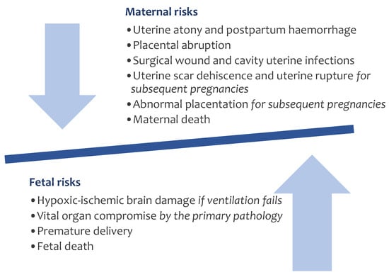
Pelvic inflammatory disease is an infectious condition affecting women’s upper genital tract, including the uterus, fallopian tubes, and ovaries. It primarily arises from an infection that spreads upward from the lower genital area. The relationship between chronic pelvic pain and coexisting conditions is a key focus in its diagnosis and treatment. This type of pain is also considered a form of reflex dystrophy, involving both neurological and psychological components, the first line treatment consists in antibiotherapy. For patients with complex or severe pelvic abscesses, surgical intervention may be considered in selected cases. The primary surgical techniques employed are open and laparoscopic surgery, both aimed for abscess removal. MRI or Doppler ultrasonography may be employed when there is a suspicion of adnexal torsion, adenomyosis or deep pelvic endometriosis, especially if the ultrasound results are unclear or inconclusive Laparoscopic surgery has increasingly become favored by both healthcare professionals and patients. Moreover, laparoscopy has emerged as the most valuable tool for diagnosing chronic pelvic pain. The approach to treating pelvic abscesses in women of reproductive age depends greatly on clinical assessments, individual patient factors, and the desire to preserve fertility. However, laparoscopy may present technical difficulties in patients with severe pelvic abscesses, particularly those with extensive adhesions or a closed-off pelvic area, requiring advanced surgical expertise. Women with associated conditions such as endometriosis often experience a more severe form of pelvic inflammatory disease, which is less responsive to antibiotics and more frequently requires surgical resolution. The surgical treatment should be performed individualized to the clinical condition of the patient and the time of intervention must be carefully chosen.
Full article


