

NCBI Bookshelf. A service of the National Library of Medicine, National Institutes of Health.
Baron S, editor. Medical Microbiology. 4th edition. Galveston (TX): University of Texas Medical Branch at Galveston; 1996.

General Concepts
Clinical Manifestations
Mycoplasma pneumoniae infection is a disease of the upper and lower respiratory tracts. Cough, fever, and headache may persist for several weeks. Convalescence is slow. Ureaplasma urealyticum infection causes nongonococcal urethritis in men, resulting in dysuria, urgency, and urethral discharge.
Structure, Classification, and Antigenic Types
Mycoplasmas are spherical to filamentous cells with no cell walls. There is an attachment organelle at the tip of filamentous M pneumoniae, M genitalium, and several other pathogenic mycoplasmas. Fried-egg-shaped colonies are seen on agar. The mycoplasmas presumably evolved by degenerative evolution from Gram-positive bacteria and are phylogenetically most closely related to some clostridia. Mycoplasmas are the smallest self-replicating organisms with the smallest genomes (a total of about 500 to 1000 genes); they are low in guanine and cytosine. Mycoplasmas are nutritionally very exacting. Many require cholesterol, a unique property among prokaryotes. Ureaplasmas require urea for growth, another unusual property. Mycoplasmas have surface antigens such as membrane proteins, lipoproteins, glycolipids, and lipoglycans. Some of the membrane proteins undergo spontaneous antigenic variation. Antibodies to surface antigens inhibit growth; various serological tests have been developed and are useful in classification.
Pathogenesis
Mycoplasmas are surface parasites of the human respiratory and urogenital tracts. Mycoplasma pneumoniae attaches to sialoglycoproteins or sialoglycolipid receptors on the tracheal epithelium via protein adhesins on the attachment organelle. The major adhesin is a 170-kilodalton (kDa) protein, named P1. Hydrogen peroxide and superoxide radicals (O2–) excreted by the attached organisms cause oxidative tissue damage. Pneumonia is induced largely by local immunologic and phagocytic responses to the parasites. Sequelae of M pneumoniae infection (mainly hematologic and neurologic) apparently have an autoimmune etiology. Several fastidious mycoplasmas may act as cofactors in activation of the aquired immunodeficiency syndrome (AIDS). Macrophage activation, cytokine induction, and superantigen properties of some mycoplasmal cell components can be considered as pathogenicity factors.
Host Defenses
IgM antibodies, followed by IgG and secretory IgA, are important in host resistance. The importance of cell-mediated immunity is unclear.
Epidemiology
Mycoplasma pneumoniae infection occurs worldwide and is more prevalent in colder months. It affects mainly children ages 5 to 9 years. It is spread by close personal contact and has a long incubation period. Ureaplasma urealyticum is spread primarily through sexual contact. Women may be asymptomatic reservoirs.
Diagnosis
Culture of M pneumoniae from sputum or a throat swab is possible, but very slow; therefore diagnosis is usually based on serologic tests. Tests using diagnostic DNA probes and amplification of specific genomic mycoplasma sequences by the polymerase-chain reaction (PCR) are being developed.
Control
There is no certified vaccine for M pneumoniae. Treatment with erythromycin or tetracyclines is effective in reducing symptoms in both M pneumoniae and U urealyticum infections.
Introduction
Mycoplasmas are the smallest and simplest self-replicating bacteria. The mycoplasma cell contains the minimum set of organelles essential for growth and replication: a plasma membrane, ribosomes, and a genome consisting of a double-stranded circular DNA molecule ( Fig. 37-1). Unlike all other prokaryotes, the mycoplasmas have no cell walls, and they are consequently placed in a separate class Mollicutes(mollis, soft; cutis, skin). The trivial term mollicutes is frequently used as a general term to describe any member of the class, replacing in this respect the older term mycoplasmas.

Figure 37-1
Electron micrograph of thin-sectioned mycoplasma cells. Cells are bounded by a single membrane showing in section the characteristic trilaminar shape. The cytoplasm contains thin threads representing sectioned chromosome and dark granules representing ribosomes. (more...)
Mycoplasmas have been nicknamed the “crabgrass” of cell cultures because their infections are persistent, frequently difficult to detect and diagnose, and difficult to cure. Contamination of cell cultures by mycoplasmas presents serious problems in research laboratories and in biotechnological industries using cell cultures. The origin of contaminating mycoplasmas is in components of the culture medium, particularly serum, or in the flora of the technician's mouth, spread by droplet infection.
Clinical Presentation
Mycoplasmal pneumonia
The term primary atypical pneumonia was coined in the early 1940s to describe pneumonias different from the typical lobar pneumonia caused by pneumococci. Several common respiratory viruses, including influenza virus and adenovirus, were shown to be responsible for a significant number of these pneumonias. From other cases, many of which developed antibodies agglutinating red blood cells in the cold (cold agglutinins), an unidentified filterable agent was isolated by Eaton and associates and was called Eaton agent. This agent was identified as a new Mycoplasma species after its successful cultivation on cell-free media in 1962. Named Mycoplasma pneumoniae, it was the first clearly documented mycoplasma pathogenic for humans.
The effects of M pneumoniae on humans include subclinical infection, upper respiratory disease, and bronchopneumonia. Most human infections do not progress to a clinically evident pneumonia. When pneumonia occurs, the onset generally is gradual and the clinical picture is one of a mild to moderately severe illness, with early complaints referable to the lower respiratory passages. Radiography frequently reveals evidence of pneumonia before physical signs are apparent. Involvement is usually limited to one of the lower lobes of the lungs, and the pneumonia is interstitial or bronchopneumonic. The course of disease varies; remittent fever, cough, and headache persist for several weeks. One of the most consistent clinical features is a long convalescence, which may extend from 4 to 6 weeks. Few fatal cases have been reported. Several unusual complications have been noted, including hemolytic anemia, polyradiculitis, encephalitis, aseptic meningitis, and central nervous system illness such as Guillain-Barré syndrome. In addition, pericarditis and pancreatitis have been observed. These sequelae may be related to the suspected immunopathology of M pneumoniae disease (see below).
Nongonococcal Urethritis and Salpingitis
Growing evidence suggests that Ureaplasma urealyticum causes nongonococcal urethritis in men free of Chlamydia trachomatis,an established agent of nongonococcal urethritis. The wide occurrence of U urealyticum in sexually active, symptom-free adults hampers research in this field. Evidence is based primarily on the production of nongonococcal urethritis symptoms in ureaplasma-free and chlamydia-free volunteers by intraurethral inoculation of U urealyticum and on a report that this disease could be cured in a chlamydia-free man only when he and his partner were treated simultanously with tetracycline, which eliminated U urealyticum from both. Ureaplasmas have also been associated with chorioamnionitis, habitual spontaneous abortion, and low-weight infants. Mycoplasma hominis, a common inhabitant of the vagina of healthy women, becomes pathogenic once it invades the internal genital organs, where it may cause pelvic inflammatory diseases such as tubo-ovarian abscess or salpingitis.
It has been suggested that Mycoplasma genitalium, isolated in 1981 from the urethral discharge of two homosexual men, may account for the tetracycline-responsive, nongonococcal urethritis cases in which chlamydias and ureaplasmas cannot be isolated (about 20 percent of all cases). However, M genitalium is so fastidious that very few clinical isolates have so far been made on the best mycoplasma medium available. Only the recent application of specific PCR amplification of the organism's DNA in clinical specimens has provided experimental proof for the relative prevalence of M genitalium in the human urogenital tract and its apparent role in male urethritis.
Mycoplasmas in AIDS and Immunocompromised Patients
The question of whether mycoplasmas act as co-factors in the development of AIDS has attracted much attention recently. Several mycoplasms have so far been incriminated: M fermentans, considered until recently a relatively rare mycoplasma of the human urogenital tract, and M penetrans , a newly-discovered human mycoplasma isolated from several AIDS patients. M pirum, a mycoplasma of an unknown host, has been recently isolated from the blood of a few AIDS patients. While, in vitro studies show that these mycoplasmas may markedly enhance pathogenicity of the human immunodeficiency virus, the possibility that the mycoplasmas may simply represent opportunistic agents found in high frequency in patients with AIDS, cannot be ruled out. Yet on the whole, with the increasing incidence of immunocompromised patients (due to AIDS, organ transplantation, etc.) evidence is accumulating for invasion of tissues and the intracellular location of some mycoplasmas, notably M fermentans and M penetrans. Extragenital infections by urogenital mycoplasmas are rather common in neonates, immunosuppressed and/or hypogammaglobulinemic patients; clinical symptoms are expressed frequently as arthritis.
Structure, Classification, and Antigenic Types
Distinguishing Properties
The coccus is the basic form of all mycoplasmas in culture. The diameter of the smallest coccus capable of reproduction is about 300 nm. In most mycoplasma cultures, elongated or filamentous forms (up to 100 μm long and about 0.4 μm thick) also occur. The filaments tend to produce truly branched mycelioid structures, hence the name mycoplasma (myces, a fungus; plasma, a form). Mycoplasmas reproduce by binary fission, but cytoplasmic division frequently may lag behind genome replication, resulting in formation of multinuclear filaments (Fig. 37-2).

Figure 37-2
Schematic presentation of the mode of mycoplasma reproduction. Cells may either divide by binary fission or first elongate to multinucleate filaments, which subsequently breakup to coccoid bodies. (From Razin S: Mycoplasmas: the smallest pathogenic procaryotes. (more...)
Some mycoplasmas possess unique attachment organelles, which are shaped as a tapered tip in M pneumoniae and M genitalium. Mycoplasma pneumoniae is a pathogen of the respiratory tract, adhering to the respiratory epithelium, primarily through the attachment organelle. Interestingly, these two human mycoplasmas exhibit gliding motility on liquid-covered surfaces. The tip structure always leads, again indicating its importance in attachment.One of the most useful distinguishing features of mycoplasmas is their peculiar fried-egg colony shape, consisting of a central zone of growth embedded in the agar and a peripheral one on the agar surface (Fig. 37-3).
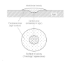
Figure 37-3
Morphology of a typical “fried-egg” mycoplasma colony.
The lack of cell walls and intracytoplasmic membranes facilitates isolation of the mycoplasma membrane in a relatively pure form. The isolated mycoplasma membrane resembles that of other prokaryotes in being composed of approximately two-thirds protein and one-third lipid. The mycoplasma lipids resemble those of other bacteria, apart from the large quantities of cholesterol in the sterol-requiring mycoplasmas.
Membrane proteins, glycolipids, and lipoglycans exposed on the cell surface are the major antigenic determinants in mycoplasmas. Antisera containing antibodies to these components inhibit growth and metabolism of the mycoplasmas and, in the presence of complement, cause lysis of the organisms. These properties are used in various serologic tests that differentiate between mycoplasma species and serotypes and detect antibodies to mycoplasmas in sera of patients (see below).
Molecular Biology
The mycoplasma genome is typically prokaryotic, consisting of a circular, double stranded DNA molecule. The Mycoplasma and Ureaplasma genomes are the smallest recorded for any self-reproducing prokaryote (Table 37-1). Therefore, there are very few genes; in some mycoplasmas the number is estimated at fewer than 500, about one sixth the number of genes in Escherichia coli. Mycoplasmas accordingly express a small number of cell proteins and lack many enzymatic activities and metabolic pathways. Their nutritional requirements are correspondingly complex, and they are dependent on a parasitic mode of life.
Table 37-1
Taxonomy and Properties of Mycoplasmas Capable of Infecting Humans a.
The dependence of mycoplasmas on their host for many nutrients explains the great difficulty of cultivation in the laboratory. The complex media for mycoplasma culture contain serum, which provides fatty acids and cholesterol for mycoplasma membrane synthesis. The requirement of most mycoplasmas for cholesterol is unique among prokaryotes. The consensus is that only a small fraction of mycoplasmas existing in nature have been cultivated so far. Some of the cultivable mycoplasmas, including the human pathogen M pneumoniae, grow very slowly, particularly on primary isolation. Ureaplasma urealyticum, a pathogen of the human urogenital tract, grows very poorly in vitro, reaching maximal titers of 107 organisms/ml of culture. Mycoplasma genitalium, another human pathogen, grows so poorly in vitro that only a few successful isolations have been achieved.
Glucose and other metabolizable carbohydrates can be used as energy sources by the fermentative mycoplasmas possessing the Embden-Meyerhof-Parnas glycolytic pathway. All mycoplasmas examined thus far possess a truncated, flavin-terminated respiratory system, which rules out oxidative phosphorylation as an ATP-generating mechanism. Breakdown of arginine by the arginine dihydrolase pathway has been proposed as a major source of ATP in nonfermentative mycoplasmas. Ureaplasmas have a requirement, unique among living organisms, for urea. Because they are non-glycolytic and lack the arginine dihydrolase pathway, it has been suggested, and later proven experimentally, that ATP is generated through an electrochemical gradient produced by ammonia liberated during the intracellular hydrolysis of urea by the organism's urease.
The mycoplasma genome is characterized by a low guanine-plus-cytosine content and by a corresponding preferential utilization of codons containing adenine and uracil, particularly in the third position. Most interesting is the use of the universal stop codon UGA as a tryptophan codon in many mycoplasmas, a rare property found so far only in mycoplasmas and in nonplant mitochondria. Resistance of mycoplasmal RNA polymerase to rifampicin is another property distinguishing mycoplasmas from the conventional eubacteria. However, apart from this resistance to rifampicin, the mycoplasmas are susceptible to antibiotics, such as tetracyclines and chloramphenicol, that inhibit protein synthesis on prokaryotic ribosomes.
Phylogeny
As the smallest and simplest self-replicating prokaryotes, the mycoplasmas pose an intriguing question: do they represent the descendents of exceedingly primitive bacteria that existed before the development of a peptidoglycan-based wall, or do they represent evolutionary degenerate eubacterial forms that have lost their cell walls? The balance of the molecular evidence, based largely on comparison of base sequences of the highly conserved ribosomal RNA (rRNA) molecules, particularly of the 16S rRNA type, favors the hypothesis of degenerative evolution. According to Woese and his colleagues, the mycoplasmas evolved as a branch of the low-guanine-plus-cytosine Gram-positive bacteria and are most closely related to two clostridia, Clostridium innocuum and C ramosum. However, the marked phenotypic and genotypic variability among mycoplasmas has led some workers to conclude that mycoplasmas evolved from a variety of walled bacteria and accordingly have a polyphyletic origin. Woese maintains that the origin of mycoplasmas is monophyletic and explains the great variety of mycoplasmas by a process of rapid evolution characteristic of the group.
Pathogenesis
All mycoplasmas cultivated and identified thus far are parasites of humans, animals, plants, or arthropods. The primary habitats of human and animal mycoplasmas are the mucous surfaces of the respiratory and urogenital tracts and the joints in some animals. Although some mycoplasmas belong to the normal flora, many species are pathogens, causing various diseases that tend to run a chronic course (Fig. 37-4).
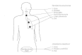
Figure 37-4
Pathogenesis and disease sites of infection by M pneumoniae and U urealyticum.
Most mycoplasmas that infect humans and other animals are surface parasites, adhering to the epithelial linings of the respiratory and urogenital tracts. Adherence is firm enough to prevent the elimination of the parasites by mucous secretions or urine. The intimate association between the adhering mycoplasmas and their host cells provides an environment in which local concentrations of toxic metabolites excreted by the parasite build up and cause tissue damage (Fig. 37-5). Moreover, because mycoplasmas lack cell walls, fusion between the membranes of the parasite and host has been suggested, and some experimental evidence for it has recently been obtained. Membrane fusion would alter the composition and permeability of the host cell membrane and enable the introduction of the parasite's hydrolytic enzymes into the host cell, events expected to cause serious damage. Recent studies have indicated the presence in mycoplasmas of antigenic variability systems. These systems, some of which are already defined in molecular genetic terms, are responsible for rapid changes in major surface protein antigens. The change in the antigenic coat of the parasite helps it to escape recognition by the immune mechanisms of the host.

Figure 37-5
Schematic presentation of a M pneumoniae organism attaching to the surface of the ciliary tracheal epithelium, as seen by electron microscopy of a thin section. The clustering of the P1 adhesin on the surface of the attachment organelle at the tip of (more...)
Because attachment of M pneumoniae and M genitalium is affected by pretreatment of the host cells with neuraminidase, sialoglycoproteins and/or sialoglycolipids of the host cell membrane appear to be receptor sites for these mycoplasmas. There is evidence that several M pneumoniae membrane proteins act as adhesins and that they have high affinity for the specific receptors for M pneumoniae on host cells. Monoclonal antibodies to one of these proteins, protein P1 (molecular weight, 170,000 daltons), inhibit attachment of the parasite. Ferritin labeling of the antibodies has shown that P1 concentrates on the tip structure of the mycoplasma, a finding that further supports the notion that the tip serves as an attachment organelle.
The results obtained with M pneumoniae were essentially duplicated recently with M genitalium and showed that in this organism, which closely resembles M pneumoniae morphologically and physiologically, a major adhesin protein, named MgPa, is clustered at the tip organelle. The genes of the major adhesins of M pneumoniae (P1) and of M genitalium (MgPa) were cloned and sequenced, allowing the characterization of these proteins. The two adhesins are alike in many respects and in fact contain extensive areas of homology, as expressed also by shared epitopes. These two proteins may be the product of an ancestral gene that underwent a horizontal gene transfer event.
The nature of the toxic factors that damage the mucosal surfaces infected by mycoplasmas is still unclear. Toxins are rarely found in mycoplasmas. Consequently, researchers considered whether the end products of mycoplasma metabolism were responsible for tissue damage. Hydrogen peroxide (H2O2), the end product of respiration in mycoplasmas, has been implicated as a major pathogenic factor ever since it was shown to be responsible for the lysis of erythrocytes by mycoplasmas in vitro; however, the production of H2O2 alone does not determine pathogenicity, as the loss of virulence in M pneumoniae is not accompanied by a decrease in H2O2 production. For the H2O2 to exert its toxic effect, the mycoplasmas must adhere closely enough to the host cell surface to maintain a toxic, steady-state concentration of H2O2 sufficient to cause direct damage, such as lipid peroxidation, to the cell membrane. The accumulation of malonyldialdehyde, an oxidation product of membrane lipids, in cells exposed to M pneumoniae supports this notion. Moreover, M pneumoniae inhibits host cell catalase by excreting superoxide radicals (O2–). This would be expected to further increase the accumulation of H2O2 at the site of parasite-host cell contact (Fig. 37-6).
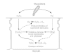
Figure 37-6
Proposed mechanism of oxidative damage to host cells by adhering M pneumoniae. by increasing concentrations of H2O2 and O2–. (Modified from Almagor M, Kahane I, Yatziv S: Role of superoxide anion in host cell injury induced by Mycoplasma pneumoniae (more...)
There is evidence that both organism-related and host-related factors are involved in the pathogenesis of mycoplasma infections. Mycoplasmas activate macrophages, and induce cytokine production and lymphocyte proliferation; the rat pathogen, Mycoplasma arthritidis, produces a potent superantigen. Thus, in the case of M pneumoniae, the host may be largely responsible for the pneumonia by mounting a local immune response to the parasite. Syrian hamsters inoculated intranasally with M pneumoniae show patchy bronchopneumonic lesions consisting of infiltration of mononuclear cells. The ablation of thymic function before the experimental infection prevents development of the characteristic pulmonary infiltration, but lengthens the period during which the organisms may be isolated from the lungs. When thymic animals are allowed to recover and then reinfected, an exaggerated and accelerated pneumonic process occurs. Epidemiologic data also suggest that repeated infections in humans are required before symptomatic disease occurs: serum antibodies to M pneumoniae can be found in most children 2 to 5 years of age, although the illness occurs with greatest frequency in individuals 5 to 15 years of age.
An immunopathologic mechanism also may explain the complications affecting organs distant from the respiratory tract in some patients infected with M pneumonia. Various autoantibodies have been detected in the sera of many of these patients, including cold agglutinins reacting with the erythrocyte I antigen, and antibodies reacting with lymphocytes, smooth muscle cells, and brain and lung antigens. Serologic cross-reactions between M pneumoniae and brain and lung antigens have been demonstrated, and these antigens are probably related to the glycolipids of M pneumoniae membranes, which are also found in most plants and in many bacteria. Clearly, host reaction varies markedly, as only about half of the patients develop cold agglutinins and complications are rare, even among individuals with anti-tissue globulins.
Host Defenses
Infection with M pneumoniae induces the development of serum antibodies that fix complement, inhibit growth of the organism and lyse the organism in the presence of complement. Generally, the first antibodies produced are of the IgM class, whereas later in convalescence the predominant antibody is IgG. Secretory IgA antibodies also develop and appear to be important in host resistance. The first infection in infancy usually is asymptomatic and generates a brief serum antibody response. Recurrent infections generate a more prolonged systemic antibody response and increasing numbers of circulating antigen-responsive lymphocytes. By late childhood, clinically apparent lower respiratory disease, including pneumonia, becomes more common. Therefore, mycoplasma respiratory disease manifestations appear to vary, depending on the state of local and systemic immunity at the time of reinfection. One hypothesis is that local immunity mediates resistance to infection and that systemic immunity contributes substantially to the pulmonary and systemic reaction characteristic of M pneumoniae pneumonia.
The relative importance of humoral and cell-mediated immunity in resistance to respiratory mycoplasma infections is still unclear. For many mycoplasma infections, such as bovine pleuropneumonia, resistance can be transferred with convalescent-phase serum, but this may not be true for all mycoplasma respiratory diseases. For example, resistance of rats to pulmonary disease induced by M pulmonis can be transferred only with spleen cells obtained from previously infected animals. Although IgA antibody may be important in resistance to mycoplasmas, other factors seem to be involved in resistance to pulmonary disease, and these factors may not be the same for all mycoplasma infections.
Epidemiology
One of the most puzzling features of M pneumoniae pneumonia is the age distribution of patients. In a survey conducted between 1964 and 1975 of more than 100,000 individuals in the Seattle area, the age-specific attack rate was highest among 5- to 9-year-old children. Rates of M pneumoniae pneumonia in the youngest age group, 0 to 4 years old, were about one-half those in school-age children, but considerably higher than in adults. Mycoplasma pneumoniae pneumonia was rarely observed in infants younger than 6 months, suggesting maternally conferred immunity (Fig. 37-7). Mycoplasma pneumoniae accounts for 8 to 15 percent of all pneumonias in young school-age children. In older children and in young adults, the organism is responsible for approximately 15 to 50 percent of all pneumonias. Infection with M pneumoniae occurs worldwide all year round but shows a predilection for the colder months, apparently because of the greater opportunity for transmission by droplet infection. Mycoplasma pneumoniae appears to require close personal contact to spread; successful spreading usually occurs in families, schools, and institutions. The incubation period ranges from 2 to 3 weeks.

Figure 37-7
Incidence of M pneumoniae pneumonia in Seattle by age, for two epidemics (1966-67 and 1974) and the endemic periods (1967-73). (From Foy HM, Kenny GE, Cooney MK, Allen ID: Long-term epidemiology of infections with Mycoplasma pneumoniae pneumonia. J Infect (more...)
Ureaplasma urealyticum is spread primarily through sexual contact. Colonization has been linked to the frequency of sexual intercourse and the number of sexual partners. Women may be asymptomatic reservoirs of infection.
Diagnosis
Culture is essential for definitive diagnosis (See below).
Culture
A routine mycoplasma medium consists of heart infusion, peptone, yeast extract, salts, glucose or arginine, and horse serum (5 to 20 percent). Fetal or newborn calf serum is preferable to horse serum. To prevent the overgrowth of the fast-growing bacteria that usually accompany mycoplasmas in clinical materials, penicillin, thallium acetate or both are added as selective agents. For Ureaplasma culture, the medium is supplemented with urea and its pH is brought to 6.0. Ureaplasm a and M genitalium are relatively sensitive to thallium, which is, therefore, omitted from their culture media. For M pneumoniae isolation, nasopharyngeal secretions are inoculated into a selective diphasic medium (pH 7.8) made of mycoplasma broth and agar and supplemented with glucose and phenol red. When M pneumoniae grows in this medium, it produces acid, causing the color of the medium to change from purple to yellow. Broth from the diphasic medium is subcultured to mycoplasma agar when a color change occurs, or at weekly intervals for a minimum of 8 weeks.
Identification
Colonies appearing on the plates can be identified as M pneumoniae by staining directly on agar with homologous fluorescein-conjugated antibody or by demonstrating that a specific antiserum to M pneumoniae inhibits their growth on agar. Colonies of ureaplasmas are usually minute (less than 100 μm in diameter); because of urea hydrolysis and ammonia liberation, the medium becomes alkaline. When manganous sulfate is added to the medium, the ureaplasma colonies stain dark brown. Isolates can be characterized in more detail by a variety of biochemical and serologic tests. More sophisticated tests, including electrophoretic analysis of cell proteins, DNA-DNA hybridization tests, mycoplasmal DNA cleavage patterns by restriction endonucleases, and PCR tests employing species-specific primers for amplification, may be performed in a research laboratory.
Serodiagnosis and Molecular Probes
Serodiagnosis consists of examining serum samples for antibodies that inhibit the growth and metabolism of the organism or fix complement with mycoplasmal antigens. A fourfold or greater rise in IgG titer is considered indicative of recent infection, whereas a sustained high antibody titer may not be significant, because a relatively high level of antibody may persist for at least 1 year after infection. A variety of rapid tests based on indirect hemagglutination of erythrocytes or latex particles coated with M pneumoniae antigens have been developed, and some are commercially available.The cold agglutinin test is less useful because only about one-half of patients develop cold agglutinins and because these antibodies also are induced by a great many other conditions.
Present techniques for laboratory diagnosis of M pneumoniae infections are of little use to the clinician because recovery by culture and identification of the mycoplasmas take at least 1 to 2 weeks. Methods for rapid laboratory diagnosis, such as direct demonstration of organisms in the respiratory specimens by nucleic acid amplification techniques, have promise but diagnostic kits are not yet commercially available.
Control
Prevention
Chemoprophylaxis of mycoplasma infections is not recommended, and no vaccine is available. Prior natural infection appears to provide the most effective resistance; however, evidence shows that M pneumoniae infections recur at intervals of several years. These observations suggest that immunity to a single natural infection is relatively short-term.
Treatment
The mycoplasmas are sensitive to tetracyclines, macrolides, and the newer quinolones, but are resistant to antibiotics that specifically inhibit bacterial cell wall synthesis. Tetracycline or erythromycin is recommended for treatment of M pneumoniae pneumonia, although effective treatment of the symptoms usually is not accompanied by eradication of the organism from the infected host. To prevent recurrence of nongonococcal urethritis caused by U urealyticum, sexual partners should be treated simultaneously with tetracycline. The incidence of tetracycline-resistant strains of U urealyticum and M hominis is on the rise.
References
- Blanchard A, Montagnier L. AIDS-associated mycoplasmas. Annu Rev Microbiol. 1994;48:687. [PubMed: 7826023]
- Cassell GH (ed) : The changing role of mycoplasmas in respiratory disease and AIDS. Clin Infect Dis 17 (Suppl 1), 1993 . [PubMed: 8104512]
- Cole BC, Griffiths MM. Triggering and exacerbation of autoimmune arthritis by the Mycoplasma arthritidis superantigen MAM. Arthritis Rheum. 1993;36:994. [PubMed: 8318046]
- Kenny GE, Kaiser GG, Cooney MK. et al. Diagnosis of Mycoplasma pneumoniae pneumonia: sensitivities and specificities of serology with lipid antigen and isolation of the organism on soy peptone medium for identification of infections. J Clin Microbiol. 1990;28:2087. [PMC free article: PMC268108] [PubMed: 2121791]
- Maniloff J, McElhaney RN, Finch LR, Baseman JB (eds): Mycoplasmas, Molecular Biology and Pathogenesis. American Society for Microbiology, Washington, 1992 .
- Pietsch K, Ehlers S, Jacobs E. Cytokine gene expression in the lungs of BALB/c mice during primary and secondary intranasal infection with Mycoplasma pneumoniae. Microbiology. 1994;140:2043. [PubMed: 7921254]
- Razin S. Molecular biology and genetics of mycoplasmas (Mollicutes). Microbiol Rev. 1985;49:419. [PMC free article: PMC373046] [PubMed: 3912655]
- Razin S. DNA probes and PCR in diagnosis of mycoplasma infections. Molec Cellular Probes. 1994;8:497. [PubMed: 7700272]
- Razin S, Barile MF (eds): The Mycoplasmas, Vol. 4, Mycoplasma Pathogenicity Academic Press, Orlando, 1985 .
- Razin S, Jacobs E. Mycoplasma adhesion. J Gen Microbiol. 1992;138:407. [PubMed: 1593256]
- Razin S, Tully JG (eds): Molecular and Diagnostic Procedures in Mycoplasmology. Academic Press, Orlando, 1995 .
- Rottem S, Kahane I (eds): Mycoplasma Cell Membranes. Plenum Press, New York, 1993 .
- Wise KS. Adaptive surface variation in mycoplasmas. Trends Microbiol. 1993;1:59. [PMC free article: PMC7133286] [PubMed: 8044463]
- The minimal cellular genome of mycoplasma.[Indian J Biochem Biophys. 1997]The minimal cellular genome of mycoplasma.Razin S. Indian J Biochem Biophys. 1997 Feb-Apr; 34(1-2):124-30.
- Review Mycoplasmas-Host Interaction: Mechanisms of Inflammation and Association with Cellular Transformation.[Microorganisms. 2020]Review Mycoplasmas-Host Interaction: Mechanisms of Inflammation and Association with Cellular Transformation.Benedetti F, Curreli S, Zella D. Microorganisms. 2020 Sep 4; 8(9). Epub 2020 Sep 4.
- Detection of genital mycoplasmas by PCR.[Methods Mol Med. 1999]Detection of genital mycoplasmas by PCR.Gilroy CB, Taylor-Robinson D. Methods Mol Med. 1999; 20:81-102.
- Review Peculiar properties of mycoplasmas: the smallest self-replicating prokaryotes.[FEMS Microbiol Lett. 1992]Review Peculiar properties of mycoplasmas: the smallest self-replicating prokaryotes.Razin S. FEMS Microbiol Lett. 1992 Dec 15; 100(1-3):423-31.
- Review [Mechanisms of genetic instability of mollicutes (mycoplasma)].[Genetika. 2001]Review [Mechanisms of genetic instability of mollicutes (mycoplasma)].Momynaliev KT, Govorun VM. Genetika. 2001 Sep; 37(9):1173-87.
- Mycoplasmas - Medical MicrobiologyMycoplasmas - Medical Microbiology
Your browsing activity is empty.
Activity recording is turned off.
See more...